. 2021 Apr 5;218(4):e20202486.
doi: 10.1084/jem.20202486.

Auto-antibodies to type I IFNs can underlie adverse reactions to yellow fever live attenuated vaccine

Affiliations

Auto-antibodies to type I IFNs can underlie adverse reactions to yellow fever live attenuated vaccine

Paul Bastard et al. J Exp Med. .

Abstract

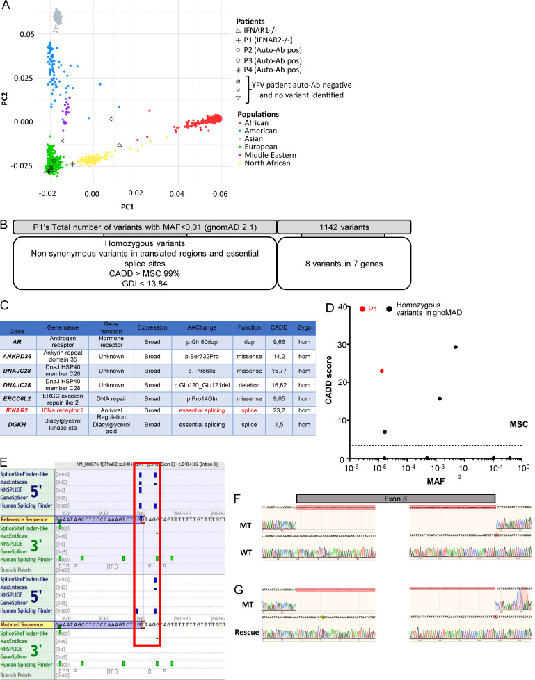
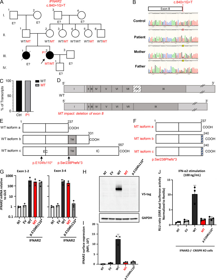
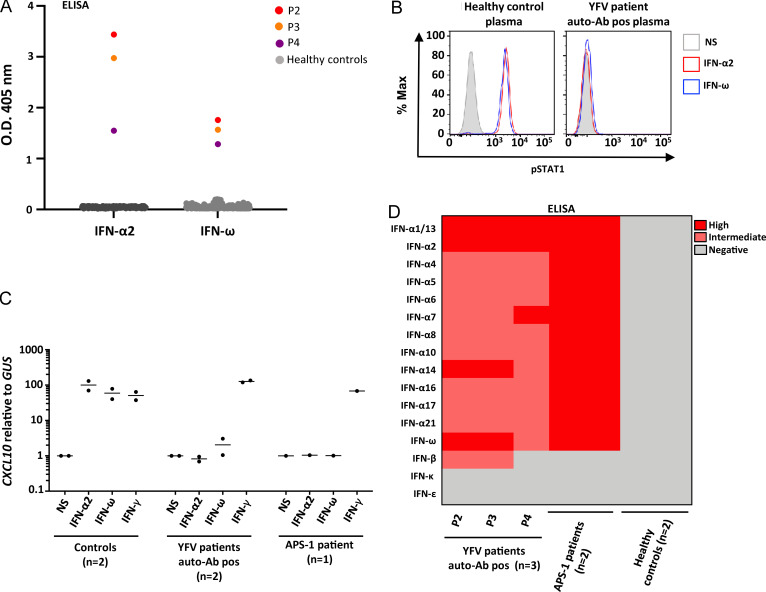
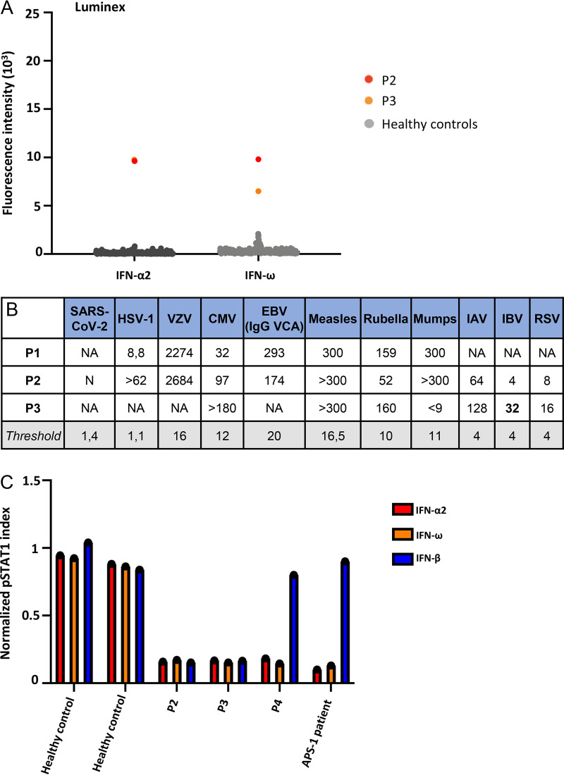
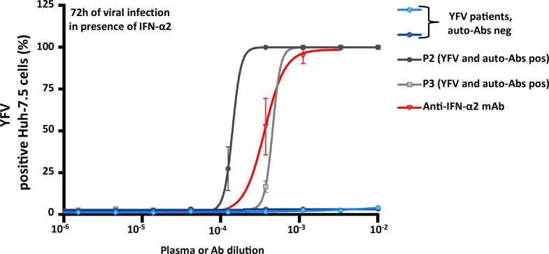
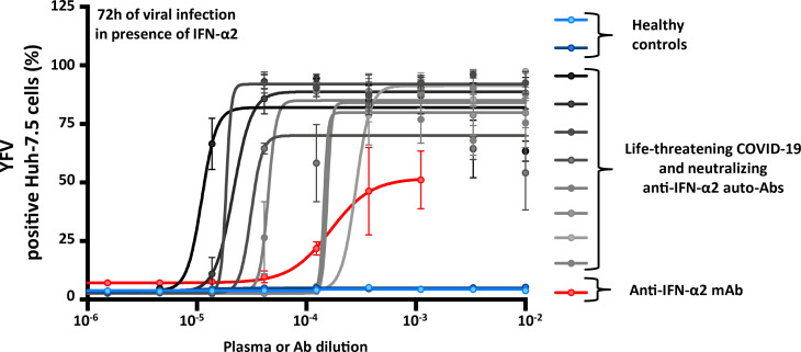
Yellow fever virus (YFV) live attenuated vaccine can, in rare cases, cause life-threatening disease, typically in patients with no previous history of severe viral illness. Autosomal recessive (AR) complete IFNAR1 deficiency was reported in one 12-yr-old patient. Here, we studied seven other previously healthy patients aged 13 to 80 yr with unexplained life-threatening YFV vaccine-associated disease. One 13-yr-old patient had AR complete IFNAR2 deficiency. Three other patients vaccinated at the ages of 47, 57, and 64 yr had high titers of circulating auto-Abs against at least 14 of the 17 individual type I IFNs. These antibodies were recently shown to underlie at least 10% of cases of life-threatening COVID-19 pneumonia. The auto-Abs were neutralizing in vitro, blocking the protective effect of IFN-α2 against YFV vaccine strains. AR IFNAR1 or IFNAR2 deficiency and neutralizing auto-Abs against type I IFNs thus accounted for more than half the cases of life-threatening YFV vaccine-associated disease studied here. Previously healthy subjects could be tested for both predispositions before anti-YFV vaccination.


Conflict of interest statement

Disclosures: A. Homma reported, "Our institution is a non-profit producer of the yellow fever vaccine. We are public institution, part of our Ministry of Health, and provide vaccine only for National Immunization Program and UNICEF, PAHO Revolving Fund, GAVI, and WHO. We are very much interested to know all relevant scientific issues involved with our vaccine." J.L. Casanova reported a patent to application number 63/055,155, filed July 22, 2020 pending. No other disclosures were reported.

Figures

Figure S1.
Figure S1.
Homozygous loss-of-function variant of IFNAR2 in a patient with YEL-AVD. (A) Principal component analysis (PCA) on the eight patients with life-threatening adverse reactions to YFV, including the patients with IFNAR1 and IFNAR2 deficiency, and those with auto-Abs against type I IFNs. (B) Filtering criteria used for the single-nucleotide variant analysis of whole-exome sequencing results for P1. (C) Single-nucleotide variants presented by the patient (P1) and meeting the filtering criteria, with the name, known function, and expression pattern of the gene. AAChange, amino acid change; Zygo, zygosity; Hom, homozygote; Dup, duplication. (D) Population genetics of homozygous coding missense IFNAR2 variants present in gnomAD. No predicted loss-of-function variants are reported in gnomAD at the homozygous state. The patient’s variant is shown in red, whereas the variants present in gnomAD are shown in black. CADD, combined annotation-dependent depletion; GDI, gene damage index; MSC, mutation significance cutoff. (E) Whole-exome sequencing results for the IFNAR2 gene in P1, as shown in ALAMUT, at the end of exon 8. The predicted splicing effect of the variant is indicated with a red square. (F) Sequencing results demonstrating complete aberrant splicing of IFNAR2 in the patient (MT) relative to the control (WT). (G) Sequencing results demonstrating normal splicing of IFNAR2 in the patient (MT) when the gDNA variant is restored to the WT form (rescue). pos, positive.
Figure 1.
Figure 1.
Homozygous loss-of-function variant of IFNAR2 in a patient with YEL-AVD. (A) Family pedigree showing the segregation of the IFNAR2 MT allele. The proband is indicated by an arrow. E?, unknown IFNAR2 genotype. (B) Sanger sequencing results for IFNAR2 for the patient, her parents, and healthy control leukocyte gDNA. (C) Exon trapping results demonstrating complete aberrant splicing in MT IFNAR2-transfected COS-7 cells. At least 100 transcripts were sequenced for the patient and the control. (D) Schematic diagram of the full-length cDNA of the WT or MT IFNAR2. The exons are numbered by roman numerals (I–IX). The 5′ and 3′ untranslated region is shown in light gray, and the coding sequences of the exons are shown in dark gray. (E) Schematic diagram of the WT IFNAR2 proteins, with the three known isoforms. The transmembrane domain is denoted “TM.” EC, extracellular domain; IC, intracellular domain. The mutation reported here is indicated in red, and the previously reported mutation is indicated in violet. (F) Schematic diagram of the MT IFNAR2 proteins, with the impact on the three known isoforms. The shaded blue area shows the three amino acids resulting from the frameshift. (G) IFNAR2 mRNA levels, determined by RT-qPCR in HEK293T cells transiently transfected with WT or MT IFNAR2 cDNA constructs; β glucuronidase (GUS) was used as an expression control. EV, empty vector; NT, nontransfected; p.E104fs110*, variant from the previously published IFNAR2−/− patient. Error bars represent standard deviation. (H) Western blot of IFNAR2 in HEK293T cells transiently transfected with IFNAR2 isoform c cDNA constructs. An Ab recognizing the V5 tag at the C-terminal end of the IFNAR2 protein was used. GAPDH was used as a loading control. A representative blot from two independent experiments is shown. (I) Graphical representation of extracellular FACS staining and the mean fluorescence intensity (MFI) of IFNAR2 in HEK293T cells transiently transfected with IFNAR2 cDNA constructs, using an Ab that recognizes the N-terminus of the protein. Cells were not permeabilized. Results representative of three independent experiments are shown. Error bars represent standard deviation. (J) Luciferase activity after IFN-α2 stimulation in IFNAR2−/− HEK293T cells generated with CRISPR/Cas-9 technology, and transiently transfected with WT or MT IFNAR2 cDNA constructs. Results shown are from three cell lines generated with three independent CRISPR guide RNAs. The bars represent the means and SEM of the results obtained with three cell lines generated with three different CRISPR guide RNAs. Each dot corresponds to the result obtained with one of these three cell lines. Ctrl, control; ISRE, interferon stimulation response element; RLU, relative light units.
Figure 2.
Figure 2.
Neutralizing auto-Abs against IFN-α2 and IFN-ω in three patients with adverse reactions to yellow fever vaccine. (A) ELISA assay for auto-Abs against IFN-α2 and IFN-ω in three patients with life-threatening adverse reactions to yellow fever vaccine and healthy controls (n = 200). (B) Representative flow cytometry plots for IFN-α2– or IFN-ω–induced pSTAT1 in healthy control cells (gated on CD14+ monocytes) in the presence of 10% healthy control plasma or anti–IFN-α2, or anti–IFN-ω auto-Ab–containing plasma from patients. Max, maximum; pos, positive; NS, not stimulated. (C) Neutralization effect on CXCL10 induction relative to GUS after the stimulation of healthy control PBMCs with IFN-α2, IFN-ω, or IFN-γ, in the presence of plasma from either healthy controls (n = 2), two patients with adverse reactions to yellow fever vaccine and auto-Abs (P2 and P3), or one APS-1 patient. (D) Auto-Abs against the different type I IFN subtypes. ELISA for auto-Abs against the 13 different IFN-α subtypes, IFN-ω, IFN-β, IFN-κ, and IFN-ε in the three patients with adverse reactions to yellow fever vaccine and auto-Abs against IFN-α2, APS-1 patients (n = 2), and healthy controls (n = 2).
Figure S2.
Figure S2.
Neutralizing auto-Abs against IFN-α2 and IFN-ω in three patients with adverse reactions to yellow fever vaccine. (A) Multiplex particle-based assay for auto-Abs against IFN-α2 and IFN-ω in two patients with life-threatening adverse reactions to yellow fever vaccine and healthy controls (n = 250; previously used in Bastard et al., 2020b). (B) Serological data for IgG Abs against various viruses, for P1, P2, and P3. The threshold value for each viral serological test is indicated. N, negative; NA, not available. In serological tests for EBV, we measured anti-VCA (viral capsid antigen) Ab levels. HSV-1, herpes simplex virus 1; VZV, varicella zoster virus; CMV, cytomegalovirus; IAV, influenza A virus; IBV, Influenza B virus; RSV, respiratory syncytial virus. (C) Normalized pSTAT1 index based on fluorescence-activated cell sorting for IFN-α2–, IFN-β–, or IFN-ω–induced pSTAT1 in healthy control cells in the presence of 10% healthy control plasma, or anti–IFN-α2, anti–IFN-β, or anti–IFN-ω auto-Ab–containing plasma from YFV patients.
Figure 3.
Figure 3.
Enhanced YFV replication, despite the presence of IFN-α2, in the presence of plasma from patients with auto-Abs against IFN-α2. YFV-17D replication, assessed 72 h after infection, in Huh-7.5 cells treated with IFN-α2 in the presence of plasma from either patients with life-threatening adverse reactions to YFV and neutralizing auto-Abs against IFN-α2 (n = 2, P2 and P3), or a commercial anti–IFN-α2 Ab, or plasma from patients with adverse events following YFV vaccination but without auto-Abs against type I IFNs (n = 2). Error bars represent SEM. neg, negative; pos, positive.
Figure S3.
Figure S3.
Enhanced YFV replication, despite the presence of IFN-α2, in the presence of plasma from patients with life-threatening COVID-19 and auto-Abs against IFN-α2. (A) YFV-17D replication, assessed 72 h after infection, in Huh-7.5 cells treated with IFN-α2, and in the presence of either plasma from patients with life-threatening COVID-19 and neutralizing auto-Abs against IFN-α2 (n = 8), or a commercial anti–IFN-α2 Ab, or plasma from healthy controls (n = 2). Error bars represent SEM.

References

    1. Adzhubei, I.A., Schmidt S., Peshkin L., Ramensky V.E., Gerasimova A., Bork P., Kondrashov A.S., and Sunyaev S.R.. 2010. A method and server for predicting damaging missense mutations. Nat. Methods. 7:248–249. 10.1038/nmeth0410-248 - DOI - PMC - PubMed
    1. Bastard, P., Manry J., Chen J., Rosain J., Seeleuthner Y., AbuZaitun O., Lorenzo L., Khan T., Hasek M., Hernandez N., et al. . 2020a. Herpes simplex encephalitis in a patient with a distinctive form of inherited IFNAR1 deficiency. J. Clin. Invest. 131:e139980. 10.1172/JCI139980 - DOI - PMC - PubMed
    1. Bastard, P., Rosen L.B., Zhang Q., Michailidis E., Hoffmann H.H., Zhang Y., Dorgham K., Philippot Q., Rosain J., Béziat V., et al. . 2020b. Autoantibodies against type I IFNs in patients with life-threatening COVID-19. Science. 370:eabd4585. 10.1126/science.abd4585 - DOI - PMC - PubMed
    1. Bousfiha, A., Jeddane L., Picard C., Al-Herz W., Ailal F., Chatila T., Cunningham-Rundles C., Etzioni A., Franco J.L., Holland S.M., et al. . 2020. Human Inborn Errors of Immunity: 2019 Update of the IUIS Phenotypical Classification. J. Clin. Immunol. 40:66–81. 10.1007/s10875-020-00758-x - DOI - PMC - PubMed
    1. Bravo García-Morato, M., Calvo Apalategi A., Bravo-Gallego L.Y., Blázquez Moreno A., Simón-Fuentes M., Garmendia J.V., Méndez Echevarría A., Del Rosal Rabes T., Domínguez-Soto Á., López-Granados E., et al. . 2019. Impaired control of multiple viral infections in a family with complete IRF9 deficiency. J. Allergy Clin. Immunol. 144:309–312.e10. 10.1016/j.jaci.2019.02.019 - DOI - PubMed

MeSH terms