. 2021 Feb 4;81(3):426-441.e8.
doi: 10.1016/j.molcel.2021.01.004.

Human DDK rescues stalled forks and counteracts checkpoint inhibition at unfired origins to complete DNA replication

Affiliations

Human DDK rescues stalled forks and counteracts checkpoint inhibition at unfired origins to complete DNA replication

Mathew J K Jones et al. Mol Cell. .

Abstract

Eukaryotic genomes replicate via spatially and temporally regulated origin firing. Cyclin-dependent kinase (CDK) and Dbf4-dependent kinase (DDK) promote origin firing, whereas the S phase checkpoint limits firing to prevent nucleotide and RPA exhaustion. We used chemical genetics to interrogate human DDK with maximum precision, dissect its relationship with the S phase checkpoint, and identify DDK substrates. We show that DDK inhibition (DDKi) leads to graded suppression of origin firing and fork arrest. S phase checkpoint inhibition rescued origin firing in DDKi cells and DDK-depleted Xenopus egg extracts. DDKi also impairs RPA loading, nascent-strand protection, and fork restart. Via quantitative phosphoproteomics, we identify the BRCA1-associated (BRCA1-A) complex subunit MERIT40 and the cohesin accessory subunit PDS5B as DDK effectors in fork protection and restart. Phosphorylation neutralizes autoinhibition mediated by intrinsically disordered regions in both substrates. Our results reveal mechanisms through which DDK controls the duplication of large vertebrate genomes.

Keywords: ATR; Cdc7; DDK; DNA replication; MERIT40; PDS5B; chemical genetics; phosphoproteomics.


Conflict of interest statement

Declaration of interests The authors declare no competing interests.

Figures

Figure 1.
Figure 1.. Human cells exhibit an escalating requirement for DDK activity across S phase.
(A) CDC7 conditional–null and analog-sensitive RPE1 cells were generated as described in STAR Methods and Figure S1. Endogenous and transgene-encoded Cdc7 were detected by immunoblotting. FLAP designates a composite localization and purification tag (FLAG-EGFP-TEV-S-peptide). (B) Cell growth in the presence or absence of 1NM-PP1 (mean ± SD; data are from three experiments). (C) Kinetics of DDK inhibition (DDKi). Lysates from Cdc7wt and Cdc7as cells treated with 1NM-PP1 were blotted to detect total or serine 40-phosphorylated MCM2. Gel loading was varied to enable comparison over a wider dynamic range. Results are representative of three experiments. (D) DDKi inhibits DNA synthesis in late S phase cells. Cdc7wt and Cdc7as cells were treated with or without 1NM-PP1 for the indicated time periods and pulse labeled with BrdU for 30 min prior to fixation. Flow cytometry was used to monitor DNA content (PI staining) and rate of synthesis (BrdU). Overlay histograms display DNA synthesis in near-4N cells (late S/G2/M phase). Results are representative of three experiments. (E) DDKi blocks progression from early to late S phase replication foci. Cdc7wt and Cdc7as cells expressing EGFP-PCNA and mKO2–hCdt1 (30–120) were synchronized in G0 via serum starvation, then refed in the presence of 1NM-PP1 and followed by spinning disk confocal microscopy. Time 0 denotes S phase entry as judged by 50% loss of mKO2-hCdt1. (F) DDKi delays formation of early replication factories. Individual traces from Cdc7wt (black) or Cdc7as (red) cells treated with 1NM-PP1 were used to compare the kinetics of mKO-hCdt1 degradation and EGFP-PCNA foci formation. (G) The number and intensity of PCNA foci in early S phase cells (t=100 to 300 min) and late S phase cells (t=400 to 1000 min) were compared using one-way ANOVA and Holm-Sidak tests. Error bars indicate SEM. Data were quantified from timelapse recordings of 8 cells per condition.
Figure 2.
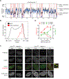
Figure 2.. DDKi causes under-replication of late regions and accumulation of stalled forks.
(A) Replication of chromosome 4 in flow-sorted RPE1 cells (black) and Cdc7as cells (blue) released from double-thymidine block in the presence of 1-NM-PP1. Significantly under-replicated regions are shaded. See also Figure S2 and Table S1. Data are from two biological replicates. (B) Distribution of under-replicated regions in DDKi cells versus genomewide replication timing (hg19). (C) Cdc7wt and Cdc7as cells were pulse labeled with EdU and chased through S phase in the presence of 1NM-PP1. At various times cells were fixed and stained with antibodies to FANCD2 (red) and 53BP1 (green). Images are maximum-intensity projections of deconvolved z stacks and are representative of three experiments. (D) Quantification of FANCD2 and 53BP1 foci in DDKi and control cells (N=3 experiments). Error bars denote SEM.
Figure 3.
Figure 3.. Replication forks require DDK to efficiently recruit RPA and activate ATR in response to nucleotide depletion.
(A) Cdc7as cells were pulse labeled with EdU (30 min), treated as specified, then fixed and stained with antibodies to FANCD2 and RPA2. Maximum-intensity projections of deconvolved z stacks and linescans are shown. Images are representative of three experiments. See also Figure S3. (B) Cdc7as cells were treated with 1NM-PP1 or 2 mM hydroxyurea (HU). ATR activation was assessed with pS345-Chk1 and pS33-RPA2 antibodies. Where indicated, gel loading was varied to enable comparison over a wider dynamic range. (C-D) HU was added to Cdc7as cells after 2 h in 1NM-PP1. (E) Cdc7wt and Cdc7as cells were treated with 1NM-PP1 with or without ATRi for the final hour of treatment. DNA-PK activation was assessed using pS4/8-RPA2 antibodies. (F) Cdc7as cells were treated with or without 1NM-PP1 for 24 h. Where indicated ATRi, RO-3306 (CDK1i), or roscovitine (CDK2i) was added for the last hour. Results are representative of three experiments.
Figure 4.
Figure 4.. S phase checkpoint inhibition induces CMG assembly, origin firing, and DNA replication in DDK-depleted Xenopus egg extracts and rescues late replication in DDKi cells.
(A) Immunodepletion of major (xDrf1) and minor (xDbf4) DDK activators from Xenopus egg extract low speed supernatants (LSS). Where indicated, lambda phosphatase was added to collapse heterogeneously phosphorylated proteins into discrete bands. (B) Sperm DNA was incubated in mock- or xDrf1/xDbf4-depleted extracts. Where indicated, caffeine was added to inhibit xATR and xATM. Chromatin pellets were analyzed by SDS-PAGE and immunoblotting. (C) Replication of sperm chromatin was monitored by [α-32P] dATP incorporation as described (Takahashi et al., 2008; Takahashi and Walter, 2005). Results from five experiments (mean ± SD) were compared using two-way ANOVA and Dunnett’s tests. (D) ATRi restores late replication in DDKi cells. Cdc7as cells were treated with 1-NM-PP1 in the presence or absence of ATRi (VE-821) or ATMi (KU-60019) for 6 h and pulse labeled with BrdU for the final 30 min. DNA content and rate of synthesis were assessed by flow cytometry. Overlay histograms compare BrdU incorporation in near-4N cells. Results are representative of three experiments. See also Figure S4. (E) Cdc7as cells were treated with or without 1NM-PP1 and ATRi for 6 h. DDK phosphorylation was assessed by immunoblotting for total or serine 40-phosphorylated MCM2. Gel loading and blot exposures were varied to facilitate comparison over a wider dynamic range.
Figure 5.
Figure 5.. Stalled forks require DDK for nascent strand protection and direct restart.
(A) Cells were pulse-labeled with IdU and CldU for 20 min each, after which extended DNA fibers were prepared and immunostained to detect individual replication tracts. Examples of initiation, elongation, and stall/termination events are shown. (B) DDKi does not affect fork velocity. Data points (n≥280 per condition) and means ± SD are from two experiments. (C-D) ATRi restores origin firing but not fork stalling in DDKi cells. Stalled/terminated forks (IdU-only tracts) and newly fired origins (CldU-only tracts) were quantified as a percentage of all replication tracts (n≥300 tracts per condition from two experiments). Means ± SEM are plotted. P-values were computed using the chi-square test. (E) Fork protection and restart assay. Cells were pulsed with IdU, treated with 1NM-PP1 and/or HU for 5 h, then washed and pulsed with CldU. Examples of fork restart, nascent strand degradation, and irreversible fork arrest are shown. (F) DDK is required for nascent strand protection. IdU tract lengths in HU-arrested Cdc7as and Cdc7wt cells (n≥300 per condition) and RPE1 cells (n≥100 per condition) were compared using a Kruskal-Wallis test. Data points and means ± SD are plotted. (G) The frequency of irreversible fork arrest/collapse after HU washout (IdU-only tracts) was determined from at least 300 tracts per condition from two experiments. Means ± SD are plotted.
Figure 6.
Figure 6.. Quantitative phosphoproteomics identifies DDK targets, including cohesin and BRCA1-A complex subunits involved in fork protection and restart.
(A) SILAC-based screen for unbiased discovery of DDK substrates and effectors. Cdc7wt and Cdc7as cells were grown in medium containing light (Arg0 and Lys0) or heavy (Arg10Lys8) amino acids, arrested in S phase with thymidine for 16 h, and treated with 1NM-PP1 for 2 h. Two biological replicates were analyzed. (B) Phosphopeptides are plotted as fold change with/without DDKi versus signal intensity. Red dots indicate phosphopeptides that were significantly and selectively downregulated by 1-NM-PP1 in Cdc7as cells in both replicates. (C) Motif analysis of high-confidence DDK-regulated phosphorylation sites (n=97). (D) The BRCA1-associated deubiquitinase (DUB) complex detects and dismantles K63-linked ubiquitin chains at DNA damage sites. DDK phosphorylation sites at the N-terminus of MERIT40 are highlighted. (E) The cohesin complex entraps chromatin fibers to mediate sister chromatid cohesion and chromatin looping. PDS5 proteins regulate the dynamics of entrapment and release via interactions with the ATPase head of Smc3, the α-kleisin Rad21/Scc1, and SA2/Scc3. PDS5B’s isoform-specific C-terminal AT hook and DDK phosphorylation sites are highlighted. (F-H) Nascent strand protection (F and G) and fork restart assays (H) were performed in wildtype, MERIT40−/−, and PDS5B−/− RPE1 cells. Where indicated, wildtype, nonphosphorylatable (4A), and IDR-deleted (ΔN, ΔC) versions of MERIT40 and PDS5B were expressed as transgenes. Data points and means ± SD are plotted and come from two experiments. See also Figure S6.
Figure 7.
Figure 7.. Vertebrate DDKs alleviate checkpoint inhibition at unfired origins and promote remodeling, protection, and restart of stalled forks.
(A) DDK promotes origin firing by counteracting the S phase checkpoint. The S phase checkpoint inhibits origin firing in the presence of replication intermediates (RIs) to avoid dNTP and RPA exhaustion. Early origins fire before checkpoint-activating RIs have accumulated and thus require minimal DDK activity. In contrast, origin firing in late S phase cells and Xenopus egg extracts is stringently repressed by the checkpoint and requires high DDK activity. (B) DDK promotes stalled fork uncoupling and RPA recruitment, which are key events in checkpoint amplification and fork protection. DDK also activates the fork protection and restart activities of cohesin and the BRCA1-A complex by phosphorylating auto-inhibitory modules in PDS5B and MERIT40.

References

    1. Abid Ali F, Renault L, Gannon J, Gahlon HL, Kotecha A, Zhou JC, Rueda D, and Costa A. (2016). Cryo-EM structures of the eukaryotic replicative helicase bound to a translocation substrate. Nature communications 7, 10708. - PMC - PubMed
    1. Alver RC, Chadha GS, Gillespie PJ, and Blow JJ (2017). Reversal of DDK-Mediated MCM Phosphorylation by Rif1-PP1 Regulates Replication Initiation and Replisome Stability Independently of ATR/Chk1. Cell reports 18, 2508–2520. - PMC - PubMed
    1. Bainor AJ, Saini S, Calderon A, Casado-Polanco R, Giner-Ramirez B, Moncada C, Cantor DJ, Ernlund A, Litovchick L, and David G. (2018). The HDAC-Associated Sin3B Protein Represses DREAM Complex Targets and Cooperates with APC/C to Promote Quiescence. Cell reports 25, 2797–2807 e2798. - PMC - PubMed
    1. Berdougo E, Terret M, and Jallepalli P. (2009). Functional dissection of mitotic regulators through gene targeting in human somatic cells. Methods Mol Biol 545, 21–37. - PubMed
    1. Berti M, Cortez D, and Lopes M. (2020). The plasticity of DNA replication forks in response to clinically relevant genotoxic stress. Nat Rev Mol Cell Biol. - PubMed

Publication types

MeSH terms