. 2021 Feb 3;13(4):585.
doi: 10.3390/cancers13040585.

Phenotypic and Proteomic Analysis Identifies Hallmarks of Blood Circulating Extracellular Vesicles in NSCLC Responders to Immune Checkpoint Inhibitors

Affiliations

Phenotypic and Proteomic Analysis Identifies Hallmarks of Blood Circulating Extracellular Vesicles in NSCLC Responders to Immune Checkpoint Inhibitors

Davide Brocco et al. Cancers (Basel). .

Abstract

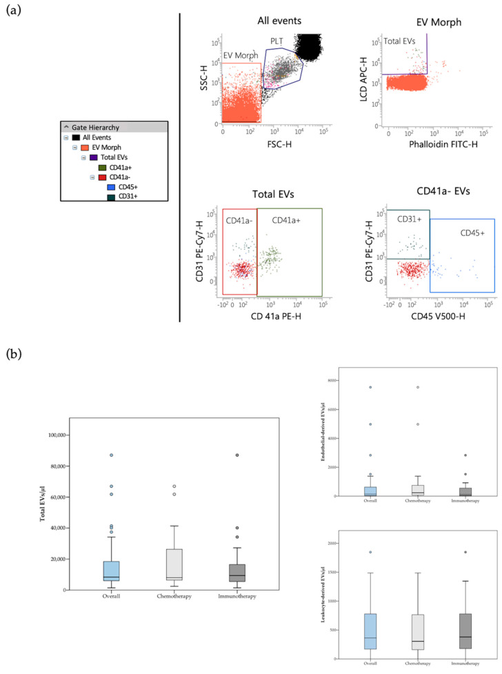
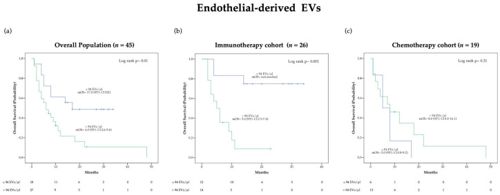
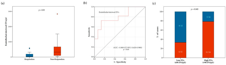
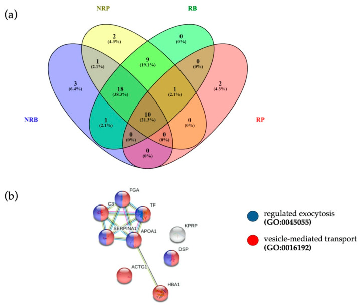
Immune checkpoint inhibitors (ICIs) induce durable clinical responses only in a subset of advanced non-small cell lung cancer (NSCLC) patients. There is a need to identify mechanisms of ICI resistance and immunotherapy biomarkers to improve clinical benefit. In this study, we evaluated the prognostic and predictive value of circulating endothelial and leukocyte-derived extracellular vesicles (EV) in patients with advanced NSCLC treated with anti-PD-1/PD-L1 agents. In addition, the relationship between total blood circulating EV proteome and response to ICIs was investigated. An optimized flow cytometry method was employed for the identification and subtyping of blood circulating EVs in 59 patients with advanced NSCLC. Blood samples were collected from patients receiving anti-PD-1/PD-L1 inhibitors (n = 31) or chemotherapy (n = 28). An exploratory proteomic analysis of sorted blood EVs was conducted in a subset of patients. Our results show that a low blood concentration of circulating endothelial-derived EVs before treatment was strongly associated to longer overall survival (p = 0.0004) and higher disease control rate (p = 0.045) in patients treated with ICIs. Interestingly, shotgun proteomics revealed that EVs of responders to anti-PD-1 therapy had a specific protein cargo before treatment. In addition, EV protein cargo was specifically modulated during immunotherapy. We identified a previously unknown association between circulating endothelial-derived extracellular vesicle concentration and immunotherapy-related clinical outcomes. We also observed differences in circulating extracellular vesicle proteome according to anti-PD-1-based treatment response in NSCLC patients. Overall, these results may contribute to the identification of novel circulating biomarkers for rational immunotherapy approaches in patients affected by NSCLC.

Keywords: biomarker; cancer immunotherapy; extracellular vesicles; immune checkpoint inhibitors; non-small cell lung cancer.


Conflict of interest statement

The authors declare no conflict of interest.

Figures

Figure 1
Figure 1
(a) Flow cytometry identification of total EVs, leukocyte-derived (CD45+ events) and endothelial-derived (CD41a−/CD31+/CD45− events) EVs in peripheral blood samples. The hierarchy of the gating strategy is represented. (b) Box plot diagram showing median blood concentration before treatment (horizontal black lines) of total EVs and EV subtypes in the overall population and in the two study groups. Extreme values are not shown.
Figure 2
Figure 2
Kaplan-Meier (KM) curves examining the relationship between overall survival and blood circulating endothelial-derived EV concentration before treatment in the overall population (a), the immunotherapy cohort (b) and the chemotherapy cohort (c).
Figure 3
Figure 3
Relationship between response to ICIs and blood circulating endothelial-derived EV concentration at treatment baseline. In panel (a), boxplot diagram showing difference in median endothelial-derived EV concentration (horizontal black lines) between responders and non-responders. Two-tailed Mann–Whitney test was performed, and p value is shown. In panel (b), receiver operating characteristic (ROC) curve illustrating predictive abilities on ICI response of blood circulating endothelial-derived EVs. In panel (c), histograms showing difference in disease control rate (DCR) according to baseline endothelial-derived EV concentration (cut-off point: 94 EVs/ μL). Blue and red bars indicate proportion of responders and non-responders, respectively. Fisher’s exact test was used to compare the two groups.
Figure 4
Figure 4
(a) Venn diagram of quantified proteins in RB, NRB, RP and NRP. (b) The 10 proteins common to four different clinical conditions were associated to “vesicle-mediated transport” (FDR = 2.27 × 10⁻⁵, red dots, PPI enrichment p-value = 9.53 × 10⁻⁷) and “regulated exocytosis” (FDR = 6.35 × 10⁻⁵, blue dots), terms coherent with EVs.
Figure 5
Figure 5
Functional Comparison Analysis between activated and inhibited pathways in sorted EVs from not responding (NR) and responding (R) patients, as compared to their respective baselines. Panel (a) highlights ROCK2 in NR and R sorted EVs compared to their baselines. Panel (b) shows IL6 modulation in NR and R EVs. Panel (c) reports the same comparison for “Chemotaxis” function. Overall ROCK2, IL6 and Chemotaxis are inhibited (blue) in responders, as compared to baseline, whereas in non-responders they result activated (orange) or unchanged (light blue). Red and green shapes represent increased or decreased measurements of identified proteins, respectively, whose fold change value is reported in the figure. Color key and symbols are reported in Supplementary Figure S8.

References

    1. Doroshow D.B., Sanmamed M.F., Hastings K., Politi K., Rimm D.L., Chen L., Melero I., Schalper K.A., Herbst R.S. Immunotherapy in Non–Small Cell Lung Cancer: Facts and Hopes. Clin. Cancer Res. 2019;25:4592–4602. doi: 10.1158/1078-0432.CCR-18-1538. - DOI - PMC - PubMed
    1. Nowicki T.S., Hu-Lieskovan S., Ribas A. Mechanisms of Resistance to PD-1 and PD-L1 Blockade. Cancer J. 2018;24:47–53. doi: 10.1097/PPO.0000000000000303. - DOI - PMC - PubMed
    1. Camidge D.R., Doebele R.C., Kerr K.M. Comparing and contrasting predictive biomarkers for immunotherapy and targeted therapy of NSCLC. Nat. Rev. Clin. Oncol. 2019;16:341–355. doi: 10.1038/s41571-019-0173-9. - DOI - PubMed
    1. Bassanelli M., Sioletic S., Martini M., Giacinti S., Viterbo A., Staddon A., Liberati F., Ceribelli A. Heterogeneity of PD-L1 Expression and Relationship with Biology of NSCLC. Anticancer Res. 2018;38:3789–3796. doi: 10.21873/anticanres.12662. - DOI - PubMed
    1. Cufaro M.C., Pieragostino D., Lanuti P., Rossi C., Cicalini I., Federici L., De Laurenzi V., Del Boccio P. Extracellular Vesicles and Their Potential Use in Monitoring Cancer Progression and Therapy: The Contribution of Proteomics. J. Oncol. 2019;2019:1639854. doi: 10.1155/2019/1639854. - DOI - PMC - PubMed

LinkOut - more resources