Review
. 2021 Feb 3;11(2):381.
doi: 10.3390/ani11020381.

Neoplasia-Associated Wasting Diseases with Economic Relevance in the Sheep Industry

Affiliations
Review

Neoplasia-Associated Wasting Diseases with Economic Relevance in the Sheep Industry

Marcelo De Las Heras et al. Animals (Basel). .

Abstract

We review three neoplastic wasting diseases affecting sheep generally recorded under common production cycles and with epidemiological and economic relevance in sheep-rearing countries: small intestinal adenocarcinoma (SIA), ovine pulmonary adenocarcinoma (OPA) and enzootic nasal adenocarcinoma (ENA). SIA is prevalent in Australia and New Zealand but present elsewhere in the world. This neoplasia is a tubular or signet-ring adenocarcinoma mainly located in the middle or distal term of the small intestine. Predisposing factors and aetiology are not known, but genetic factors or environmental carcinogens may be involved. OPA is a contagious lung cancer caused by jaagsiekte sheep retrovirus (JSRV) and has been reported in most sheep-rearing countries, resulting in significant economic losses. The disease is clinically characterized by a chronic respiratory process as a consequence of the development of lung adenocarcinoma. Diagnosis is based on the detection of JSRV in the tumour lesion by immunohistochemistry and PCR. In vivo diagnosis may be difficult, mainly in preclinical cases. ENA is a neoplasia of glands of the nasal mucosa and is associated with enzootic nasal tumour virus 1 (ENTV-1), which is similar to JSRV. ENA enzootically occurs in many countries of the world with the exception of Australia and New Zealand. The pathology associated with this neoplasia corresponds with a space occupying lesion histologically characterized as a low-grade adenocarcinoma. The combination of PCR and immunohistochemistry for diagnosis is advised.

Keywords: enzootic nasal adenocarcinoma; enzootic nasal tumour virus; jaagsiekte sheep retrovirus; ovine pulmonary adenocarcinoma; small intestinal adenocarcinoma.


Conflict of interest statement

The authors declare no conflict of interest.

Figures

Figure 1
Figure 1
Gross pathology and histopathology of the small intestinal adenocarcinoma. (A) Sheep 5 years old. Serous surface on distal third segment of the small intestine. Firm and white mass extending over serosa of convoluted intestinal segment. Plaited whitish cords are extended on the mesentery. (B) Sheep 5 years old. Section of the intestine showing single polypoid pedunculated mass causing stricture. Lumen of the intestine greatly dilated is observed proximal to the mass. (C) Histopathology of an intestinal sample from A. Acinar and tubular structures of various sizes lined by cuboidal or columnar epithelial cells replace the intestinal mucosa. Small groups or cords of epithelial cells are also seen. Moderate desmoplastic reaction together with lymphocyte infiltration is observed. Hematoxylin-eosin. 200×. (D) Histopathology of an intestinal sample from B. Signet ring cells characterized by mucin-filled cytoplasm; peripheralized crescent-shaped nuclei are numerous in this area of the tumour. Hematoxylin-eosin. 100×.
Figure 2
Figure 2
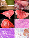
Ovine pulmonary adenocarcinoma (OPA): clinical signs and pathology. (A) Four year old sheep showing OPA clinical signs. When head is lowered, frothy sero-mucous fluid comes from the nostrils and pours over the floor. (B) Post-mortem examination of thoracic cavity. Classical OPA form. Left lung with cranio-ventral areas purple in colour and consolidated. The rest of the lung shows an increase in volume and is lighter in colour. (C) Right caudal lung section. Classical OPA. Lung section showing granular appearance foci and foamy fluid pouring from main airways. (D) Left lung from a sheep showing atypical OPA form. Multiple white nodules of various sizes distributed throughout the lung surface. (E) Section of lung from right caudal lobe. Atypical OPA. Multiple white nodules with various sizes: bigger ones coalescing close to a central main bronchus and others expanding to close lung areas. (F) Histopathology of OPA lesion. Alveolar epithelial cells proliferate following a lepidic pattern. Alveolar wall is replaced by cuboidal cells into a tubular or papiliform pattern. Tumour stroma is infiltrated mainly by lymphocytes. Numerous macrophages are filling alveolar lumens in the tumours and in the close areas. Hematoxylin-eosin. 100×. (G) Histopathology of OPA lesion. Bronchiole with epithelium transformed into papiliform growth almost completely filling the lumen. Hematoxylin-Eosin. 100×. (H) Immunohistochemistry using a mouse monoclonal antijaagsiekte sheep retrovirus envelope (JSRV Env) protein. Cellular membranes and cytoplasms of neoplastic cells of a tumour nodule are labelled. In the adjacent alveoli, positive cells are observed either isolated or in small groups. 100×.
Figure 3
Figure 3
Enzootic nasal adenocarcinoma (ENA) clinical signs and pathology. (A) Sheep 3 years old. Right nasal nostril showing sero-mucous fluid discharge. As a consequence, right nostril looks cleaner and less hairy (washed nose). (B) Sheep 4 years old. Sagittal section of the skull showing replacement of normal ethmoidal turbinate by a multinodular or polypoid mass covered by mucus pressing close structures. (C) Histopathology of ENA. Epithelial cells proliferate into acinar and tubular patterns in inner parts; meanwhile, external zones show a predominant papillary pattern. Hematoxylin-Eosin. 100×. (D) Histological section of ENA. Immunohistochemistry. Monoclonal antibody against JSRV Env protein. Positive material is almost exclusively located on the apical membranes of both types of growths. 100×.

Similar articles

Cited by

References

    1. Head K.W., Munro R. Tumours. In: Martin W.B., Aitken I.D., editors. Diseases of Sheep. 3rd ed. Blackwell Science; Oxford, UK: 2000. pp. 381–386.
    1. Else R. Tumors of Sheep. In: Aitken I.D., editor. Diseases of Sheep. 4th ed. Blackwell Publishing; Oxford, UK: 2007. pp. 443–448.
    1. Monot M., Archer F., Gomes M., Mornex J.F., Leroux C. Advances in the study of transmissible respiratory tumours in small ruminants. Vet. Microbiol. 2015;181:170–177. doi: 10.1016/j.vetmic.2015.08.008. - DOI - PubMed
    1. Munday J.S., Brennan M.M., Jaber A.M., Kiupel M. Ovine intestinal adenocarcinomas: Histologic and phenotypic comparison with human colon cancer. Comp. Med. 2006;56:136–141. - PubMed
    1. Gray M.E., Sullivan P., Marland J.R.K., Greenhalgh S.N., Meehan J., Gregson R., Clutton R.E., Cousens C., Griffiths D.J., Murray A., et al. Novel Translational Ovine Pulmonary Adenocarcinoma Model for Human Lung Cancer. Front. Oncol. 2019;9:534. doi: 10.3389/fonc.2019.00534. - DOI - PMC - PubMed

LinkOut - more resources