

Identifying a Role of Red and White Wine Extracts in Counteracting Skin Aging: Effects of Antioxidants on Fibroblast Behavior
- PMID: 33546215
- PMCID: PMC7913355
- DOI: 10.3390/antiox10020227
Identifying a Role of Red and White Wine Extracts in Counteracting Skin Aging: Effects of Antioxidants on Fibroblast Behavior
Abstract
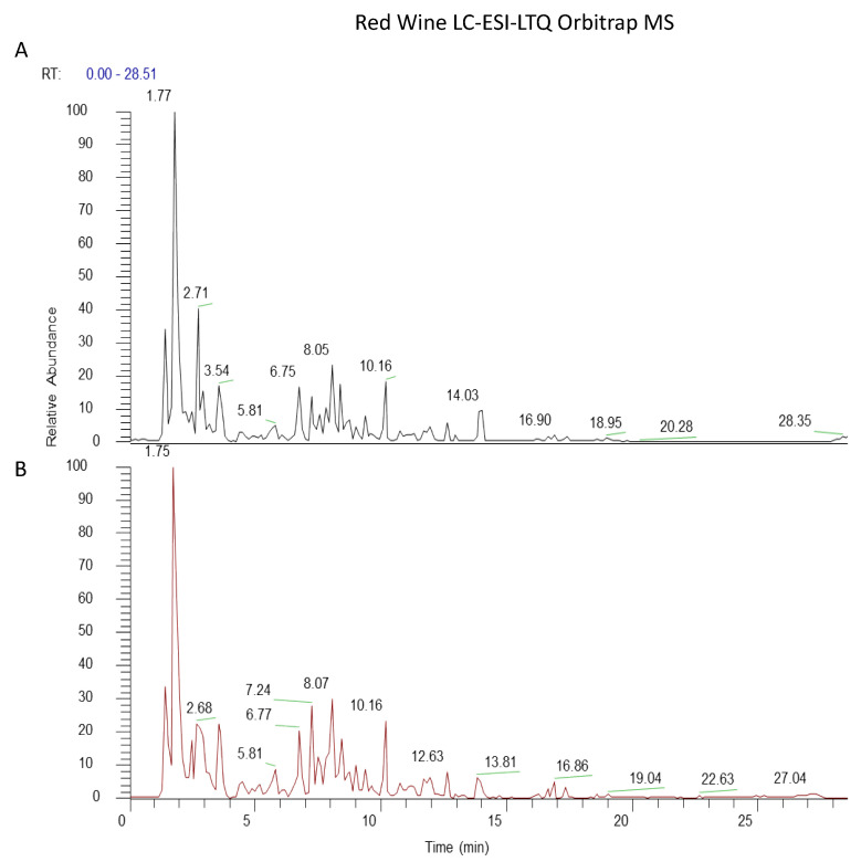
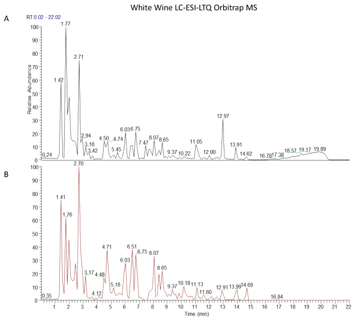
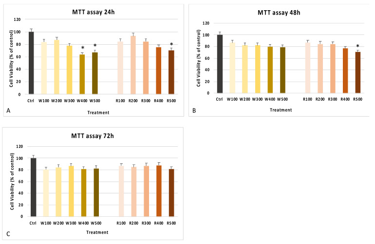
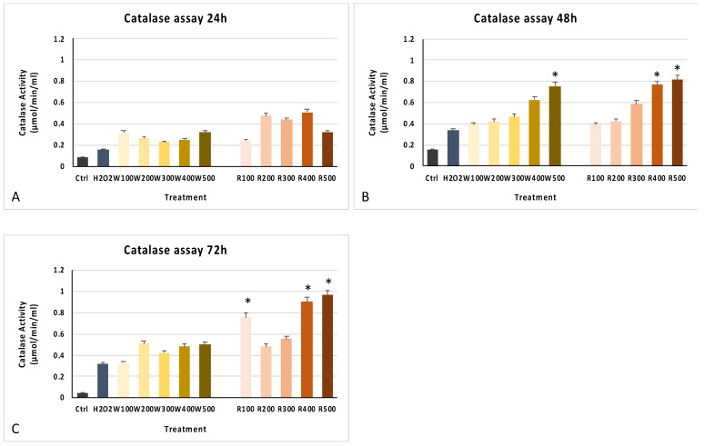
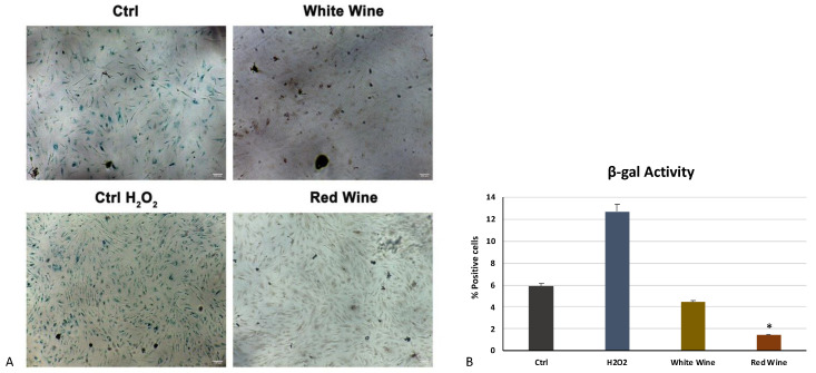
Dermal fibroblasts are the main actor in many proteins' secretion, including collagen, preserving skin function. Free radicals are involved in skin aging and damages involving different cellular components. The imbalance between reactive oxygen species (ROS) amount and natural antioxidant enzymes negatively affects skin homeostasis. Natural compounds have recently emerged as a potential anti-aging tool in tissue regeneration. In the present paper we evaluated the antioxidant activity of white and red wines, considering their probable use, as raw materials, for the formulation of cosmetic products with anti-aging properties. We studied a method that would allow the removal of the alcoholic fraction of wines and determined their composition by LC-MS analysis. We then tested the possible cytotoxic effects of red and white wines on fibroblasts by 3-(4,5-dimethylthiazol-2-yl)-2,5-diphenyltetrazolium (MTT) assay, and their antioxidant activity by the catalase activity test in stressing conditions. Finally, we evaluated their anti-aging potential through the β-galactosidase colorimetric assay. Our results showed that wine extracts exhibit a remarkable antioxidant and anti-aging activity, especially on cells exposed to a marked stressful event. These properties could suggest their possible application as cosmetical products for skin regeneration.
Keywords: antioxidants; bioactive molecules; cell proliferation; cell senescence; oxidative stress; skin aging.
Conflict of interest statement
The authors declare no conflict of interest.
Figures
Similar articles
-
23-Hydroxytormentic acid protects human dermal fibroblasts by attenuating UVA-induced oxidative stress.Photodermatol Photoimmunol Photomed. 2017 Mar;33(2):92-100. doi: 10.1111/phpp.12294. Epub 2017 Feb 9. Photodermatol Photoimmunol Photomed. 2017. PMID: 28106292
-
DNA macroarray study of skin aging-related genes expression modulation by antioxidant plant extracts on a replicative senescence model of human dermal fibroblasts.Phytother Res. 2011 May;25(5):686-93. doi: 10.1002/ptr.3308. Epub 2010 Nov 11. Phytother Res. 2011. PMID: 21077257
-
Ultraviolet-B Protective Effect of Flavonoids from Eugenia caryophylata on Human Dermal Fibroblast Cells.Pharmacogn Mag. 2015 Oct;11(Suppl 3):S397-406. doi: 10.4103/0973-1296.168979. Pharmacogn Mag. 2015. PMID: 26929573 Free PMC article.
-
Skin aging and oxidative stress: Equol's anti-aging effects via biochemical and molecular mechanisms.Ageing Res Rev. 2016 Nov;31:36-54. doi: 10.1016/j.arr.2016.08.001. Epub 2016 Aug 9. Ageing Res Rev. 2016. PMID: 27521253 Review.
-
The study of phenolic compounds as natural antioxidants in wine.Crit Rev Food Sci Nutr. 2003;43(3):233-44. doi: 10.1080/10408690390826509. Crit Rev Food Sci Nutr. 2003. PMID: 12822671 Review.
Cited by
-
Protective Effects of Mogroside V on Oxidative Stress Induced by H2O2 in Skin Fibroblasts.Drug Des Devel Ther. 2021 Dec 1;15:4901-4909. doi: 10.2147/DDDT.S337524. eCollection 2021. Drug Des Devel Ther. 2021. PMID: 34880600 Free PMC article.
-
Antioxidant Supplements versus Health Benefits of Brief/Intermittent Exposure to Potentially Toxic Physical or Chemical Agents.Curr Issues Mol Biol. 2021 Jul 10;43(2):650-664. doi: 10.3390/cimb43020047. Curr Issues Mol Biol. 2021. PMID: 34287292 Free PMC article. Review.
References
-
- Fore J. A review of skin and the effects of aging on skin structure and function. Ostomy Wound Manag. 2006;52:24–35. - PubMed
LinkOut - more resources
Full Text Sources
Other Literature Sources
