. 2021 Feb 3;13(4):606.
doi: 10.3390/cancers13040606.

The Oxygen-Generating Calcium Peroxide-Modified Magnetic Nanoparticles Attenuate Hypoxia-Induced Chemoresistance in Triple-Negative Breast Cancer

Affiliations

The Oxygen-Generating Calcium Peroxide-Modified Magnetic Nanoparticles Attenuate Hypoxia-Induced Chemoresistance in Triple-Negative Breast Cancer

Fong-Yu Cheng et al. Cancers (Basel). .

Abstract

Cancer response to chemotherapy is regulated not only by intrinsic sensitivity of cancer cells but also by tumor microenvironment. Tumor hypoxia, a condition of low oxygen level in solid tumors, is known to increase the resistance of cancer cells to chemotherapy. Triple-negative breast cancer (TNBC) is the most aggressive subtype of breast cancer. Due to lack of target in TNBC, chemotherapy is the only approved systemic treatment. We evaluated the effect of hypoxia on chemotherapy resistance in TNBC in a series of in vitro and in vivo experiments. Furthermore, we synthesized the calcium peroxide-modified magnetic nanoparticles (CaO2-MNPs) with the function of oxygen generation to improve and enhance the therapeutic efficiency of doxorubicin treatment in the hypoxia microenvironment of TNBC. The results of gene set enrichment analysis (GSEA) software showed that the hypoxia and autophagy gene sets are significantly enriched in TNBC patients. We found that the chemical hypoxia stabilized the expression of hypoxia-inducible factor 1α (HIF-1α) protein and increased doxorubicin resistance in TNBC cells. Moreover, hypoxia inhibited the induction of apoptosis and autophagy by doxorubicin. In addition, CaO2-MNPs promoted ubiquitination and protein degradation of HIF-1α. Furthermore, CaO2-MNPs inhibited autophagy and induced apoptosis in TNBC cells. Our animal studies with an orthotopic mouse model showed that CaO2-MNPs in combination with doxorubicin exhibited a stronger tumor-suppressive effect on TNBC, compared to the doxorubicin treatment alone. Our findings suggest that combined with CaO2-MNPs and doxorubicin attenuates HIF-1α expression to improve the efficiency of chemotherapy in TNBC.

Keywords: autophagy; chemoresistance; hypoxia; nanocarriers; triple-negative breast cancer.


Conflict of interest statement

The authors declare no conflict of interest.

Figures

Figure 1
Figure 1
Gene set enrichment analysis (GSEA) of hypoxia and autophagy gene expressions in primary tumors and normal tissues derived from patients with triple-negative breast cancer (TNBC). All of the expressed genes were uploaded into GSEA for enrichment analysis. The Harris hypoxia (A) and Gene Ontology (GO) regulation of autophagy (B) gene sets database were used as the gene set collection for analysis. NES, normalized enrichment score; NOM p-val, nominal p value.
Figure 2
Figure 2
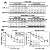
The chemical hypoxia (CoCl2) stabilizes hypoxia-inducible factor 1α (HIF-1α) protein and causes doxorubicin (DOX) resistance in TNBC cells. (A) the protein level of HIF-1α in 4T1 and MDA-MB-231 cells treated with CoCl2 for 24 and 48 h. (B) effect of DOX on cell viability of 4T1 and MDA-MB-231 cells in the absence or presence of CoCl2. Cells were pretreated with CoCl2 for 48 h and then treated with DOX for 24 h. * p < 0.05 DOX alone compared with 100 μM CoCl2 + DOX. # p < 0.05 DOX alone compared with 200 μM CoCl2 + DOX.
Figure 3
Figure 3
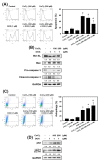
Detection of apoptosis and autophagy in 4T1 cells that received DOX and/or CoCl2 treatments. (A) apoptosis was analyzed by flow cytometry with an Annexin V apoptosis detection kit. 4T1 cells were pretreated with CoCl2 for 24 h and then treated with DOX for 24 h. * p < 0.05 compared with control. # p < 0.05 DOX alone compared with 200 μM CoCl2 + DOX. (B) effects of apoptosis-related protein expression in 4T1 cells treated with DOX and CoCl2 alone or in combination. Cells were pretreated with CoCl2 for 24 h and then treated with DOX (1 μM) for 24 h. (C) measurement of acidic vesicular organelles (AVOs) in acridine orange (AO)-stained cells using flow cytometry. 4T1 cells were pretreated with CoCl2 for 24 h and then treated with DOX for 24 h. * p < 0.05 compared with control. # p < 0.05 DOX alone compared with 200 μM CoCl2 + DOX. (D) effects of autophagy-related protein expression in 4T1 cells treated with DOX and CoCl2 alone or in combination. Cells were pretreated with CoCl2 for 24 h and then treated with DOX (1 μM) for 24 h.
Figure 4
Figure 4
Chemical and physical characteristics of calcium peroxide-modified magnetic nanoparticles (CaO2-MNPs). (A) TEM images of COOH-modified MNPs (COOH-MNPs) and CaO2-MNPs. The used solvents for preparing TEM samples of COOH-MNPs and CaO2-MNPs were water and polyethylene glycol (M.W. 200) (PEG200), respectively. (B) dissolved oxygen kinetics after the addition of Ca2+/COOH-MNPs or CaO2-MNPs (0.1 mL; Fe concentration: 0.9 mg/mL) into 20 mL of water at room temperature. (C) optical photographs of Ca2+/COOH-MNPs and CaO2-MNPs incubated with water. (D) the pH value profiles after addition of Ca2+/COOH-MNPs and CaO2-MNPs (0.1 mL; Fe concentration: 0.9 mg/mL) in 20 mL of PBS buffer (pH 7.4) or water at room temperature.
Figure 5
Figure 5
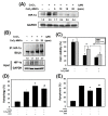
CaO2-modified MNPs cause protein degradation and ubiquitination of hypoxia-inducible factor 1α (HIF-1α), and reverts the effects of DOX on apoptosis and autophagy in 4T1 cells. (A) Western blot analysis for HIF-1α protein expression. Cells were incubated with CoCl2 (100 μM) for 24 h, treated with CaO2-MNPs for 24 h, then harvested for protein extraction and then subjected to western blot analysis. (B) immunoprecipitation (IP) assay for HIF-1α ubiquitination. 4T1 cells incubated with CoCl2 (100 μM) for 24 h and treated with CaO2-MNPs for 24 h. The total protein lysates harvested from 4T1 cells were immunoprecipitated with anti-HIF-1α antibody. The complexes and the input were examined using western blot analysis with anti-ubiquitin antibody. Effect of CaO2-MNPs on cell viability (C), autophagy (D) and apoptosis (E) of 4T1 cells. Cells were pretreated with CoCl2 (100 μM) for 8 h, treated with CaO2-MNPs (20 ppm) for 16 h, then incubated with DOX (1 μM) for 24 h. * p < 0.05 DOX alone compared with DOX + CoCl2. # p < 0.05 DOX + CoCl2 compared with DOX + CoCl2 + CaO2-MNPs.
Figure 6
Figure 6
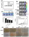
The anti-cancer effects of DOX in combination with CaO2-MNPs in an orthotopic mouse model. The female Balb/c mice were injected with 5 × 10⁴ 4T1 cells orthotopically into the 4th mammary fat pad. (A) the body weight was measured weekly in the different groups. (B) the bioluminescence imaging of tumors was used in vivo imaging system (IVIS) 200 system with Living Image Software. (C) the tumor weight was analyzed after sacrificing. * p < 0.05 control compared with DOX + CaO2-MNPs. (D) specific immunostaining was conducted on 4T1 tumor sections showing the expression of HIF-1α and LC3 in response to various treatments in vivo. Scale bars = 50 µm.
Figure 7
Figure 7
CaO2-MNPs attenuate hypoxia-induced chemoresistance in TNBC. CaO2-MNPs release oxygen and degrade HIF-1α protein by ubiquitination. Hypoxia inhibit the DOX-induced apoptosis and DOX-increased autophagy in TNBC cells. CaO2-MNPs promote cell death against DOX-induced resistance under hypoxia through the inhibition of autophagy and induction of apoptosis. Therefore, co-treatment with CaO2-MNPs and DOX could be an effective approach to attenuate HIF-1α-mediated hypoxic responses to modulate TNBC. The figure was created with BioRender.com.

References

    1. Kumar P., Aggarwal R. An overview of triple-negative breast cancer. Arch. Gynecol. Obstet. 2016;293:247–269. doi: 10.1007/s00404-015-3859-y. - DOI - PubMed
    1. Shen M., Jiang Y.Z., Wei Y., Ell B., Sheng X., Esposito M., Kang J., Hang X., Zheng H., Rowicki M., et al. Tinagl1 suppresses triple-negative breast cancer progression and metastasis by simultaneously inhibiting integrin/FAK and EGFR signaling. Cancer Cell. 2019;35:64–80. doi: 10.1016/j.ccell.2018.11.016. - DOI - PubMed
    1. Cheng X., He P., Lee T., Yao H., Li R., Shen Y. High activities of BACE1 in brains with mild cognitive impairment. Am. J. Pathol. 2014;184:141–147. doi: 10.1016/j.ajpath.2013.10.002. - DOI - PMC - PubMed
    1. Marotti J.D., de Abreu F.B., Wells W.A., Tsongalis G.J. Triple-negative breast cancer: Next-Generation sequencing for target identification. Am. J. Pathol. 2017;187:2133–2138. doi: 10.1016/j.ajpath.2017.05.018. - DOI - PubMed
    1. Piret J.P., Cosse J.P., Ninane N., Raes M., Michiels C. Hypoxia protects HepG2 cells against etoposide-induced apoptosis via a HIF-1-independent pathway. Exp. Cell. Res. 2006;312:2908–2920. doi: 10.1016/j.yexcr.2006.05.018. - DOI - PubMed

LinkOut - more resources