Review
. 2021 Feb 3;18(4):1415.
doi: 10.3390/ijerph18041415.

Neurological Sequelae in Patients with COVID-19: A Histopathological Perspective

Affiliations
Review

Neurological Sequelae in Patients with COVID-19: A Histopathological Perspective

Francesco Fisicaro et al. Int J Environ Res Public Health. .

Abstract

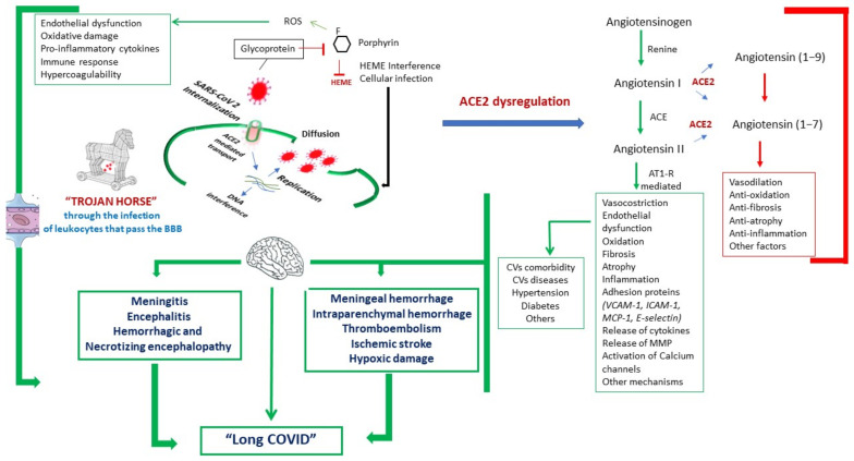
Background: Neuroinvasive properties of SARS-CoV-2 have allowed the hypothesis of several pathogenic mechanisms related to acute and chronic neurological sequelae. However, neuropathological correlates have been poorly systematically investigated, being retrieved from reports of single case or limited case series still.

Methods: A PubMed search was carried out to review all publications on autopsy in subjects with "COronaVIrus Disease-19" (COVID-19). Among them, we focused on histological findings of the brain, which were compared with those from the authors' autoptic studies performed in some COVID-19 patients.

Results: Only seven studies reported histological evidence of brain pathology in patients deceased for COVID-19, including three with reverse transcription-quantitative polymerase chain reaction evidence of viral infection. All these studies, in line with our experience, showed vascular-related and infection-related secondary inflammatory tissue damage due to an abnormal immune response. It is still unclear, however, whether these findings are the effect of a direct viral pathology or rather reflect a non-specific consequence of cardiovascular and pulmonary disease on the brain.

Conclusions: Notwithstanding the limited evidence available and the heterogeneity of the studies, we provide a preliminary description of the relationship between SARS-CoV-2 and brain sequelae. Systematic autoptic investigations are needed for accurate detection and adequate management of these patients.

Keywords: COVID-19; SARS-CoV-2; autopsy; histopathology; long-term prognosis; neuroinvasion; neuropathology; outcome; pathogenesis.


Conflict of interest statement

The authors declare no conflict of interest.

Figures

Figure 1
Figure 1
Main pathophysiological mechanisms proposed in patients with COronaVIrus Disease-19 (COVID-19) and related acute and long-term neurological manifestations. Angiotensin-converting enzyme-2 (ACE2) and iron chelated with any porphyrin, irrespective of the valence state of the iron atom (HEME) dysregulation, as well as direct action on vascular endothelium, immune response, and inflammation (with the subsequent release of inflammatory mediators), and the “Trojan horse hypothesis” (i.e., leukocytes-carriers of infection through the blood–brain barrier) have been proposed as the main mechanisms responsible for brain lesions. The inhibitory pathways are shown in red, while the activatory pathways are indicated in green, along with morphological changes responsible for cardiovascular and brain diseases, especially in comorbid patients. Legend: ACE2: angiotensin converting enzyme-2; AT1-R: angiotensin II receptor type; BBB: blood–brain barrier; CVs: cardiovascular manifestations; HEME: iron chelated with any porphyrin, irrespective of the valence state of the iron atom; ICAM-1: intercellular adhesion molecule 1; MCP-1: monocyte chemoattractant protein-1; MMP: matrix metalloproteinase; ROS: reactive oxygen species; VCAM-1: vascular cell adhesion molecule 1.
Figure 2
Figure 2
Main anatomopathological findings from authors’ autoptic studies carried out in some COVID-19 patients. (AD): vascular-related changes; (A): meningeal erythrocyte extravasations (arrows) [Hematoxylin and eosin (H&E) stain ×10]; (B): focal and multiple intraparenchymal erythrocyte extravasations (arrows) (H&E stain ×40); (C): fibrinoid material aggregated in the vessel lumen (thrombo-embolus), with associated pale parenchymal areas as from ischemic damage in the first phase (arrows) (H&E stain ×10); (D): initial transformation of neuronal cytoplasm and nucleus with the presence of pink neurons from hypoxia (arrows) (H&E stain ×40). (EH): infection-related changes (H&E stain ×20); (E): inflammatory lymphomonocyte infiltrate in meningitis (arrows); (F): area of inflammatory infiltrate with neuronal destruction and reactive aspects (arrows); (G,H): combined aspects that are often associated in hemorrhagic (G) and necrotizing (H) encephalopathy, characterized by erythrocyte extravasations, mainly in perivascular regions, with areas of adjacent intraparenchymal necrosis and more or less evident inflammatory rate (arrows). (I,J): normal brain findings (H&E stain ×10); (I): cerebral cortex, near a sulcus; (J): cerebral cortex, near the hippocampus.

References

    1. Baig A.M., Khaleeq A., Ali U., Syeda H. Evidence of the COVID-19 Virus Targeting the CNS: Tissue Distribution, Host-Virus Interaction, and Proposed Neurotropic Mechanisms. ACS Chem. Neurosci. 2020;11:995–998. doi: 10.1021/acschemneuro.0c00122. - DOI - PubMed
    1. Baig A.M., Sanders E.C. Potential Neuroinvasive Pathways of SARS-CoV-2: Deciphering the Spectrum of Neurological Deficit Seen in Coronavirus Disease-2019 (COVID-19) J. Med. Virol. 2020;92:1845–1857. doi: 10.1002/jmv.26105. - DOI - PMC - PubMed
    1. Moriguchi T., Harii N., Goto J., Harada D., Sugawara H., Takamino J., Ueno M., Sakata H., Kondo K., Myose N., et al. A First Case of Meningitis/Encephalitis Associated with SARS-Coronavirus-2. Int. J. Infect. Dis. 2020;94:55–58. doi: 10.1016/j.ijid.2020.03.062. - DOI - PMC - PubMed
    1. Bernard-Valnet R., Pizzarotti B., Anichini A., Demars Y., Russo E., Schmidhauser M., Cerutti-Sola J., Rossetti A.O., Du Pasquier R. Two Patients with Acute Meningoencephalitis Concomitant with SARS-CoV-2 Infection. Eur. J. Neurol. 2020;27:e43–e44. doi: 10.1111/ene.14298. - DOI - PMC - PubMed
    1. Ye M., Ren Y., Lv T. Encephalitis as a Clinical Manifestation of COVID-19. Brain. Behav. Immun. 2020;88:945–946. doi: 10.1016/j.bbi.2020.04.017. - DOI - PMC - PubMed

LinkOut - more resources