. 2021 Feb 3;13(2):241.
doi: 10.3390/v13020241.

Mucosal-Associated Invariant T (MAIT) Cells Are Highly Activated and Functionally Impaired in COVID-19 Patients

Affiliations

Mucosal-Associated Invariant T (MAIT) Cells Are Highly Activated and Functionally Impaired in COVID-19 Patients

Sebastian Deschler et al. Viruses. .

Abstract

Coronavirus disease 2019 (COVID-19), caused by infection with severe acute respiratory syndrome coronavirus 2 (SARS-CoV-2), comprises mild courses of disease as well as progression to severe disease, characterised by lung and other organ failure. The immune system is considered to play a crucial role for the pathogenesis of COVID-19, although especially the contribution of innate-like T cells remains poorly understood. Here, we analysed the phenotype and function of mucosal-associated invariant T (MAIT) cells, innate-like T cells with potent antimicrobial effector function, in patients with mild and severe COVID-19 by multicolour flow cytometry. Our data indicate that MAIT cells are highly activated in patients with COVID-19, irrespective of the course of disease, and express high levels of proinflammatory cytokines such as IL-17A and TNFα ex vivo. Of note, expression of the activation marker HLA-DR positively correlated with SAPS II score, a measure of disease severity. Upon MAIT cell-specific in vitro stimulation, MAIT cells however failed to upregulate expression of the cytokines IL-17A and TNFα, as well as cytolytic proteins, that is, granzyme B and perforin. Thus, our data point towards an altered cytokine expression profile alongside an impaired antibacterial and antiviral function of MAIT cells in COVID-19 and thereby contribute to the understanding of COVID-19 immunopathogenesis.

Keywords: COVID-19; SARS-CoV-2; mucosal-associated invariant T (MAIT) cells.


Conflict of interest statement

The authors declare no conflict of interest with regards to the present manuscript.

Figures
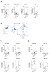
Figure 1
Figure 1
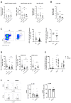
Alterations in adaptive immune cell frequency in COVID-19 patients. (A) Frequency of conventional T cells (HC n = 15; COVID mild n = 17; COVID severe n = 9), B cells (HC n = 15; COVID mild n = 10; COVID severe n = 9) and Treg cells (HC n = 10; COVID mild n = 9; COVID severe n = 8) in peripheral blood of COVID-19 patients and healthy controls (HC); (B) Flow cytometry gating strategy for identification of T cell subsets; Frequency of CD4+ (C) and CD8+ (D) T cell subsets in peripheral blood of COVID-19 patients and healthy controls (HC n = 10; COVID mild n = 9; COVID severe n = 8). Data are presented as mean ± SEM and were pooled from three independent experiments; each symbol represents one patient; ** p < 0.01, *** p < 0.001, **** p < 0.0001 vs. HC or as indicated, data were assessed using one-way analysis of variance (ANOVA) with Tukey’s multiple comparisons test, ns = not significant; Tcm= central memory T cells, Tem = effector memory T cells and Tte = terminally differentiated T effector cells.
Figure 2
Figure 2
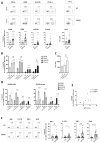
MAIT cells are significantly and continuously reduced in peripheral blood of COVID-19 patients, regardless of disease severity. (A) Frequency of natural killer (NK) cells ( HC n = 12; COVID mild n = 9; COVID severe n = 10), NKT-like cells (HC n = 13, COVID mild n = 12; COVID severe n = 10) and (B) γδ2 T cells (HC n = 12; COVID mild n = 12; COVID severe n = 10) in peripheral blood of COVID-19 patients and healthy controls (HC); (C,D) mucosal-associated invariant T (MAIT) cell frequency (C: HC n = 26 or 12; COVID n = 38 or 20; D: HC n = 26; COVID mild total n = 22, acute n = 13, convalescent n = 9; COVID severe total n = 17, acute n = 6, convalescent n = 11), (E) T cell receptor (TCR) co-receptor expression (HC n = 16; COVID n = 26) and (F) IL-12 receptor (IL-12R): HC n = 12; COVID n = 20) and IL-18 receptor (IL-18R): HC n = 12; COVID n = 22) expression in COVID-19 patients and HC. Data are presented as mean ± SEM and were pooled from 3 (A,B) or >10 (C,D) independent experiments; each symbol represents one patient; * p < 0.05, ** p < 0.01, *** p < 0.001 and **** p < 0.0001 vs. HC or as indicated; data were assessed using one-way analysis of variance (ANOVA) with Tukey’s multiple comparisons test (A,B,D), unpaired t-test (C,F) or two-way ANOVA with Sidak’s multiple comparisons test (E). ns = not significant; DN = CD4/CD8 double-negative.
Figure 3
Figure 3
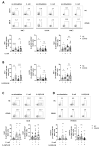
MAIT cells of COVID-19 patients express high levels of activation markers and cytokines ex vivo. (AD) Ex vivo expression of surface activation markers in MAIT cells from peripheral blood of COVID-19 patients and healthy controls (HC); (E) Spearman correlation between expression of HLA-DR in MAIT cells in peripheral blood of COVID-19 patients and SAPS II score at day of sampling; (F) Ex vivo intracellular cytokine expression in MAIT cells from peripheral blood of COVID-19 patients and HC, representative data from one patient and summary data; (A,E,F) each symbol represents one patient. (A) CD38: HC n = 18 and COVID n = 30; CD69: HC n = 21 and COVID n = 38; HLA-DR: HC n = 21 and COVID = 35; CTLA-4: HC n = 20 and COVID n = 36; PD-1: HC n = 23 and COVID n = 34. (B) HC n = 13, COVID mild n = 22 and COVID severe n = 18; (C) HC n = 26, COVID mild n = 17 and COVID severe n = 9; (D) HC n = 19, COVID acute n = 13 and COVID convalescent n = 9; (E) n = 8; (F) IFNγ: HC n = 19 and COVID n = 33; IL-17A: HC n = 23 and COVID n = 33; TNFα: HC n = 23 and COVID n = 34; GrzB: HC n = 23 and COVID n = 34. Data are presented as mean ± SEM and were pooled from six (AD) or four (E) independent experiments; * p < 0.05, ** p < 0.01, *** p < 0.001 and **** p < 0.0001 vs. HC or as indicated. Data were assessed using unpaired t-test (A,F), two-way analysis of variance (ANOVA) with Tukey’s multiple comparisons test (B,D) or one-way ANOVA with Tukey’s multiple comparisons test (C). ns = not significant.
Figure 4
Figure 4
MAIT cells from peripheral blood of COVID-19 patients show an altered cytokine expression profile. Intracellular cytokine expression in MAIT cells in a PBMC pool from peripheral blood of COVID-19 patients and healthy controls (HC) following co-culture with E. coli at 10 bacteria per cell (A,C,D) or stimulation with 50 ng/mL IL-12/IL-18 (B,C,D) for 24 h. Data are presented as mean ± SEM and were pooled from three independent experiments; each symbol represents one patient. (A) IFNγ: HC n = 16 and COVID n = 22; IL-17A/TNFα: HC n = 20 and COVID n = 24; (B) IFNγ: HC n = 16 and COVID n = 21; IL-17A/TNFα: HC n = 26 and COVID n = 20; (C,D) HC n = 16 and COVID n = 21; * p < 0.05 and ** p < 0.01 vs. unstimulated control. Data were assessed using paired t-test (A,B) or one-way analysis of variance (ANOVA) with Dunnett’s multiple comparisons test (C,D). ns = not significant.

References

    1. Chen N., Zhou M., Dong X., Qu J., Gong F., Han Y., Qiu Y., Wang J., Liu Y., Wei Y., et al. Epidemiological and clinical characteristics of 99 cases of 2019 novel coronavirus pneumonia in Wuhan, China: A descriptive study. Lancet. 2020;395:507–513. doi: 10.1016/S0140-6736(20)30211-7. - DOI - PMC - PubMed
    1. Huang C., Wang Y., Li X., Ren L., Zhao J., Hu Y., Zhang L., Fan G., Xu J., Gu X., et al. Clinical features of patients infected with 2019 novel coronavirus in Wuhan, China. Lancet. 2020;395:497–506. doi: 10.1016/S0140-6736(20)30183-5. - DOI - PMC - PubMed
    1. The epidemiological characteristics of an outbreak of 2019 novel coronavirus diseases (COVID-19) in China. Zhonghua Liu Xing Bing Xue Za Zhi. 2020;41:145–151. - PubMed
    1. Guan W.-J., Ni Z.-Y., Hu Y., Liang W.-H., Ou C.-Q., He J.-X., Liu L., Shan H., Lei C.-L., Hui D.S.C., et al. Clinical Characteristics of Coronavirus Disease 2019 in China. N. Engl. J. Med. 2020;382:1708–1720. doi: 10.1056/NEJMoa2002032. - DOI - PMC - PubMed
    1. Verity R., Okell L.C., Dorigatti I., Winskill P., Whittaker C., Imai N., Cuomo-Dannenburg G., Thompson H., Walker P.G.T., Fu H., et al. Estimates of the severity of coronavirus disease 2019: A model-based analysis. Lancet Infect. Dis. 2020;20:669–677. doi: 10.1016/S1473-3099(20)30243-7. - DOI - PMC - PubMed

Publication types