. 2021 Feb 5;19(1):38.
doi: 10.1186/s12951-021-00776-w.

Anti-bacterial and wound healing-promoting effects of zinc ferrite nanoparticles

Affiliations

Anti-bacterial and wound healing-promoting effects of zinc ferrite nanoparticles

Reihaneh Haghniaz et al. J Nanobiotechnology. .

Abstract

Background: Increasing antibiotic resistance continues to focus on research into the discovery of novel antimicrobial agents. Due to its antimicrobial and wound healing-promoting activity, metal nanoparticles have attracted attention for dermatological applications. This study is designed to investigate the scope and bactericidal potential of zinc ferrite nanoparticles (ZnFe2O4 NPs), and the mechanism of anti-bacterial action along with cytocompatibility, hemocompatibility, and wound healing properties.

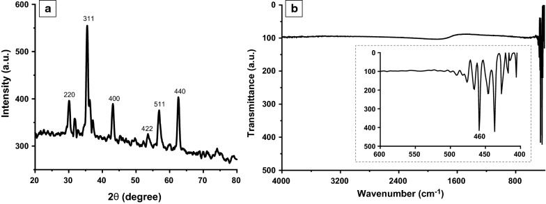
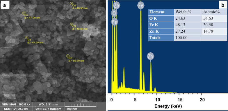
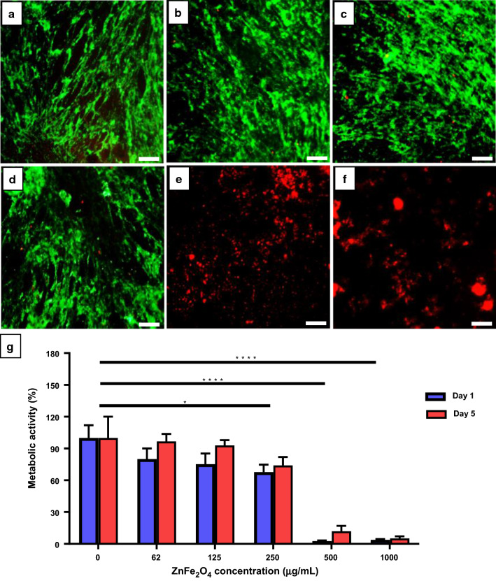
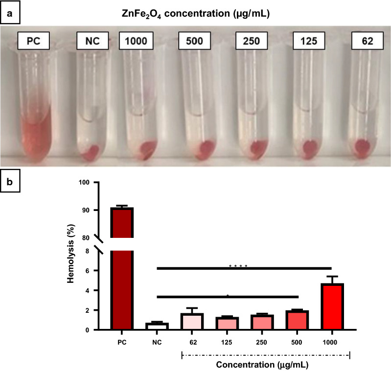
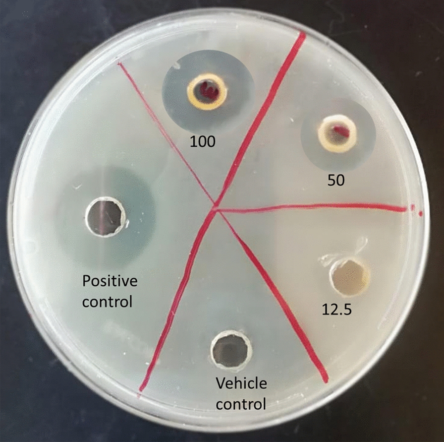
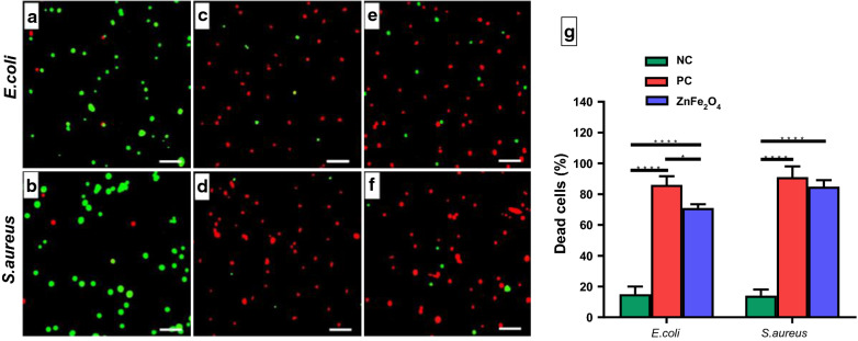
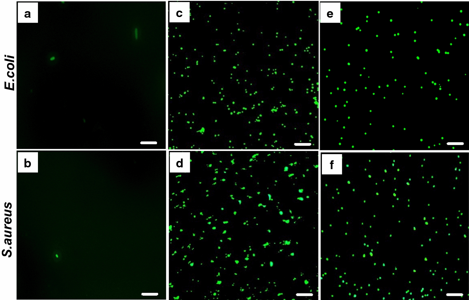
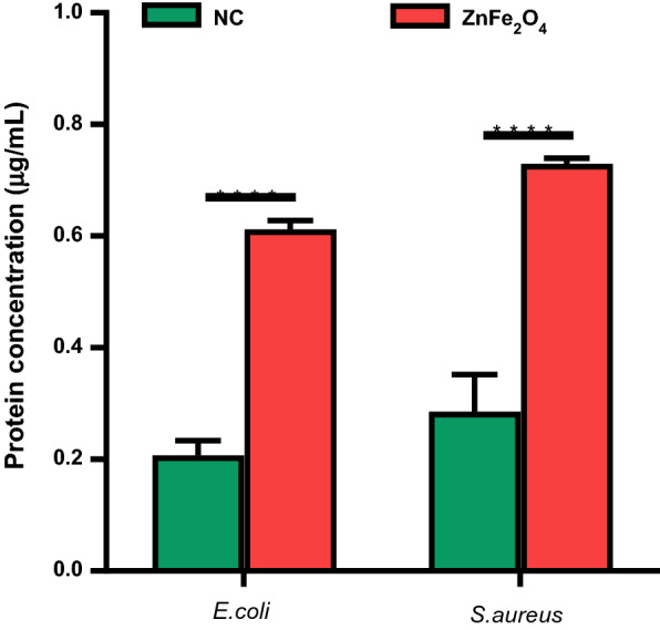
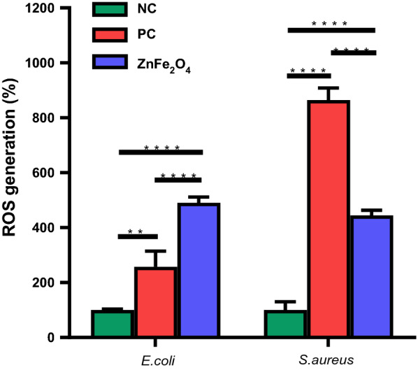
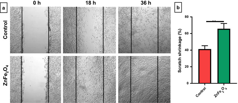
Results: ZnFe2O4 NPs were synthesized via a modified co-precipitation method. Structure, size, morphology, and elemental compositions of ZnFe2O4 NPs were analyzed using X-ray diffraction pattern, Fourier transform infrared spectroscopy, and field emission scanning electron microscopy coupled with energy-dispersive X-ray spectroscopy. In PrestoBlue and live/dead assays, ZnFe2O4 NPs exhibited dose-dependent cytotoxic effects on human dermal fibroblasts. In addition, the hemocompatibility assay revealed that the NPs do not significantly rupture red blood cells up to a dose of 1000 µg/mL. Bacterial live/dead imaging and zone of inhibition analysis demonstrated that ZnFe2O4 NPs showed dose-dependent bactericidal activities in various strains of Gram-negative and Gram-positive bacteria. Interestingly, NPs showed antimicrobial activity through multiple mechanisms, such as cell membrane damage, protein leakage, and reactive oxygen species generation, and were more effective against gram-positive bacteria. Furthermore, in vitro scratch assay revealed that ZnFe2O4 NPs improved cell migration and proliferation of cells, with noticeable shrinkage of the artificial wound model.

Conclusions: This study indicated that ZnFe2O4 NPs have the potential to be used as a future antimicrobial and wound healing drug.

Keywords: Antibiotics; Antimicrobial activity; Biocompatibility; Hemocompatibility; Nanoparticles; Spinel ferrites; Wound healing; Zinc ferrites.


Conflict of interest statement

The authors declare that they have no competing interests.

Figures

Fig. 1
Fig. 1
a X-ray diffraction (XRD) patterns, b Fourier-transform infrared spectroscopy (FTIR) spectrum of the synthesized zinc ferrite (ZnFe2O4) NPs
Fig. 2
Fig. 2
a Field-emission scanning electron microscopy (FE-SEM) image of zinc ferrite nanoparticles (ZnFe2O4 NPs) exhibits spherical particles with an average size of 47.9 ± 2.5 nm. b Energy-dispersive X-ray spectroscopy (EDX) spectrum of ZnFe2O4 NPs shows the elemental compositions reflecting with the determined weight percentage and atomic percentage of all elements (i.e., zinc, Zn; iron, Fe; and oxygen, O) according to stoichiometry
Fig. 3
Fig. 3
In vitro biocompatibility assay. Representative fluorescence microscopy images of human dermal fibroblast cells (HDF) stained with ethidium homodimer-1 (red color; dead cells) and calcein-AM (green color; live cells) after 5 days incubation with ZnFe2O4 NPs at concentrations of a 0 µg/mL (control), b 62 µg/mL, c 125 µg/mL, d 250 µg/mL, e 500 µg/mL, and f 1000 µg/mL. Scale bars show 500 µm. g The percent (%) metabolic activity of HDF cells on day 1 and 5 after subjecting to different concentrations of ZnFe2O4 NPs. Error bars denote the standard deviations of 4 replicates, and * represents the significance of the reduction in % metabolic activities of treated cells as compared to control. The data were analyzed by two-way ANOVA, and *p  0.05, ****p  0.001 were considered statistically significant
Fig. 4
Fig. 4
Hemocompatibility of zinc ferrite nanoparticles (ZnFe2O4 NPs). a Photographs of hemolysis assay to detect the presence of hemoglobin in the supernatant of ZnFe2O4 NPs treated samples. b Hemolysis percentage of ZnFe2O4 NPs treated samples versus positive and negative control. PEG and Triton X-100 lysed blood cells served as a negative control (NC) and positive control (PC), respectively. The values presented in the graph are mean ± SD of triplicate and *p  0.05, ****p  0.0001 were considered statistically significant
Fig. 5
Fig. 5
Bacterial growth inhibition of zinc ferrite nanoparticles (ZnFe2O4 NPs). Representative zone of inhibition images after treatment of S. aureus bacteria with different concentrations of ZnFe2O4 NPs (12.5–100 µg/mL) compared to the vehicle control (DMSO) and positive control (1 % silver sulfadiazine cream)
Fig. 6
Fig. 6
Bacterial live/dead assay. Fluorescence micrographs of E. coli and S. aureus after treatment with 100 µg/mL of ZnFe2O4 NPs. a Negative control E. coli cells without any treatment. b S. aureus negative control without treatment. c E. coli treated with tetracycline (positive control). d S. aureus treated with tetracycline (positive control). e E. coli treated with ZnFe2O4 NPs (100 µg/mL). f S. aureus treated with ZnFe2O4 NPs (100 µg/mL). g Percent (%) dead cells of E. coli and S. aureus after ZnFe2O4 NPs treatment as compared to the negative control (NC) and positive control (PC). Experiments were performed in triplicate. The percent dead cells of E. coli and S. aureus were significantly higher in nanoparticle-treated groups and positive control as compared to the negative control. *p ≤ 0.05 and ****p ≤ 0.0001 are statistically significant. Scale bars show 200 µm
Fig. 7
Fig. 7
Membrane permeability assay. Fluorescence images of E. coli and S. aureus showing the influx of FITC after treatment with zinc ferrite (ZnFe2O4) NPs and tetracycline (positive control). a Untreated E. coli (negative control). b Untreated S. aureus (negative control). c E. coli treated with tetracycline (positive control). d S. aureus treated with tetracycline (positive control). e E. coli treated with 100 µg/mL of ZnFe2O4 NPs. f S. aureus treated with 100 µg/mL of ZnFe2O4 NPs. Tetracycline and ZnFe2O4 NPs induced membrane damage to both Gram-positive and Gram-negative bacteria, resulted in the membrane permeability to the green fluorescent FITC dye. Scale bars show 100 µm
Fig. 8
Fig. 8
Protein leakage assay. The concentration of protein detected in ZnFe2O4 NPs (100 µg/mL) treated E. coli and S. aureus after 8 h of treatment versus untreated bacteria as a negative control (NC). All experiments were done in triplicate, and the data are presented as mean ± SD. ****p ≤ 0.0001 was considered statistically significant
Fig. 9
Fig. 9
Reactive oxygen species (ROS) assay. Intracellular ROS production after 8 h of treatment with ZnFe2O4 NPs (100 µg/mL) in E. coli and S. aureus. Hydrogen peroxide (H2O2) treated cells were taken as a positive control (PC), and bacteria without treatment served as a negative control (NC). All experiments were performed in triplicates, and data are presented as mean ± SD. **p ≤ 0.01 and****p ≤ 0.0001 were considered as statistically significant
Fig. 10
Fig. 10
Scratch assay of NIH-3T3 fibroblast cells in the presence of ZnFe2O4 NPs (100 µg/mL). a Bright-field images of the negative control (untreated cells) and ZnFe2O4 NPs treated cells after 0, 18, and 36 h of incubation. b Percent (%) scratch shrinkage of ZnFe2O4 NPs treated cells versus the untreated control after 18 h. The experiments were performed in triplicates, and data are presented as mean ± SD. The data were analyzed using t-test, where **p ≤ 0.01 was considered statistically significant

References

    1. Blair JM, et al. Molecular mechanisms of antibiotic resistance. Nat Rev Microbiol. 2015;13(1):42–51. doi: 10.1038/nrmicro3380. - DOI - PubMed
    1. Frieri M, Kumar K, Boutin A. Antibiotic resistance. J Infect Public Health. 2017;10(4):369–78. doi: 10.1016/j.jiph.2016.08.007. - DOI - PubMed
    1. Saravanan M, Ramachandran B, Barabadi H. The prevalence and drug resistance pattern of extended spectrum β–lactamases (ESBLs) producing Enterobacteriaceae in Africa. Microb Pathog. 2018;114:180–92. doi: 10.1016/j.micpath.2017.11.061. - DOI - PubMed
    1. Mohamed H. One year prevalence of critically ill burn wound bacterial infections in surgical ICU in Egypt: Retrospective study. Egyptian Journal of Anaesthesia. 2016;32(3):431–4. doi: 10.1016/j.egja.2016.01.005. - DOI
    1. Church D, et al. Burn wound infections. Clin Microbiol Rev. 2006;19(2):403–34. doi: 10.1128/CMR.19.2.403-434.2006. - DOI - PMC - PubMed

MeSH terms