. 2021 Feb 5;7(6):eabe3362.
doi: 10.1126/sciadv.abe3362. Print 2021 Feb.

The fibrinolytic system enables the onset of Plasmodium infection in the mosquito vector and the mammalian host

Affiliations

The fibrinolytic system enables the onset of Plasmodium infection in the mosquito vector and the mammalian host

Thiago Luiz Alves E Silva et al. Sci Adv. .

Abstract

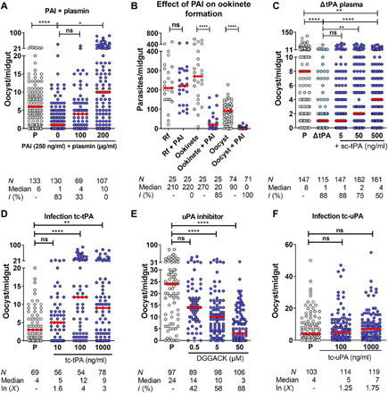
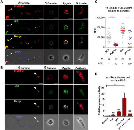
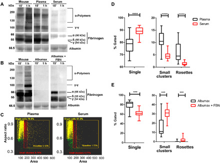
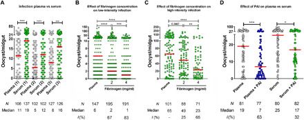
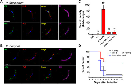
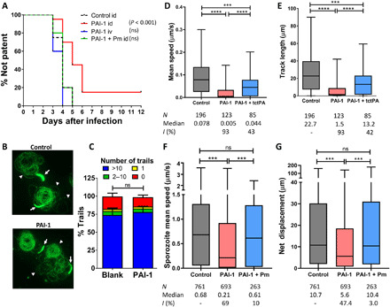
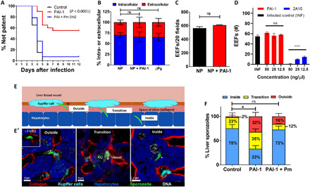
Plasmodium parasites must migrate across proteinaceous matrices to infect the mosquito and vertebrate hosts. Plasmin, a mammalian serine protease, degrades extracellular matrix proteins allowing cell migration through tissues. We report that Plasmodium gametes recruit human plasminogen to their surface where it is processed into plasmin by corecruited plasminogen activators. Inhibition of plasminogen activation arrests parasite development early during sexual reproduction, before ookinete formation. We show that increased fibrinogen and fibrin in the blood bolus, which are natural substrates of plasmin, inversely correlate with parasite infectivity of the mosquito. Furthermore, we show that sporozoites, the parasite form transmitted by the mosquito to humans, also bind plasminogen and plasminogen activators on their surface, where plasminogen is activated into plasmin. Surface-bound plasmin promotes sporozoite transmission by facilitating parasite migration across the extracellular matrices of the dermis and of the liver. The fibrinolytic system is a potential target to hamper Plasmodium transmission.


Figures

Fig. 1
Fig. 1. Plasminogen activators are required for P. falciparum mosquito infection.
An. gambiae mosquitoes were fed on infected blood reconstituted with human plasma and supplemented with the indicated inhibitors, agonists, or phosphate-buffered saline (PBS). Mosquito midguts were dissected 10 days later, and oocysts were counted. (A) PAI-1 inhibits oocyst formation in An. gambiae mosquitoes. Plasmin supplementation reverses the PAI-1 inhibition. Data pooled from three independent experiments. (B) PAI-1 (2.5 μg/ml) does not inhibit gametogenesis (round form formation) but inhibits ookinete formation. Round forms (Rf; female gametes plus zygotes) and ookinetes were isolated from the midgut blood bolus and counted at 22 to 23 hours and oocysts at 10 days after infection. Data are pooled from three independent experiments. (C) tPA-depleted plasma (ΔtPA) negatively affects oocyst development and supplementation with single-chain tPA (sc-tPA; the pro-enzyme) partially rescues the inhibition. Data pooled from four independent experiments. (D) Addition of active tc-tPA to an infectious blood meal increases oocyst development. Data pooled from three independent experiments. (E) The uPA inhibitor DGGACK reduces oocyst development. Data are pooled from two independent experiments. (F) Addition of active two-chain uPA (tc-uPA) to an infectious blood meal does not increase oocyst development. Data pooled from four independent experiments. N, number of midguts; I (%), percent inhibition; In (X), fold increase; ΔtPA, tPA-depleted plasma; P, plasma; red horizontal lines, medians. Kruskal-Wallis with Dunn’s posttest. *P < 0.05, **P < 0.01, ****P < 0.0001; ns, not significant.
Fig. 2
Fig. 2. Plasminogen, tPA, and uPA bind to the surface of P. falciparum sexual stages.
(A and B) Plasminogen (PLG), tPA (A), and uPA (B) bind to male (♂) and female (♀) gametes, zygotes, and ookinetes. Immunofluorescence images merged with phase contrast images in the bottom row. White arrowheads point to localization of plasminogen and plasminogen activators on the heads of microgametes. Scale bars for male gametes, 2 μm; scale bars for female gametes, zygotes and ookinetes, 6 μm. (C) The lysine analog tranexamic acid (TA) inhibits tPA and PLG association with the parasite surface. Purified female gametes were incubated with PLG and tPA in the presence or absence of TA. Plasminogen and tPA binding were determined by IFA with an anti-PLG and an anti-tPA antibody. Data pooled from two independent experiments. N, number of midguts; I (%), percent inhibition; RFU, relative fluorescence units. (Mann-Whitney U test, ****P < 0.0001). (D) tPA activates plasminogen on the surface of female gametes. Female gametes were incubated with the sc-tPA proenzyme. After binding, the cells were washed and incubated with PLG. Plasmin activity was measured using a chromogenic substrate. Controls: Gametes were incubated with either PBS, PLG, sc-tPA alone, or with sc-tPA followed by PLG in the presence of TA. Error bars represent SEM from three independent experiments. Analysis of variance (ANOVA) followed by Tukey’s multiple comparison test. **P < 0.01.
Fig. 3
Fig. 3. Detection of fibrin and RBC aggregates in mosquito midguts.
(A and B) Western blot showing the detection of fibrin in the blood bolus of mosquitoes fed directly on a mouse or human RBCs reconstituted with (A) plasma or serum or (B) Albumax or Albumax plus fibrinogen (FBN; 4 mg/ml). Anti-human fibrinogen antibody detects fibrin—γ-γ dimers (95 kDa) and α-polymers (>120 kDa). Albumin from the Ponceau stained membrane was used as loading control. (C) Representative ImageStream flow cytometry plots of RBC aggregation in one mosquito midgut blood bolus (related to fig. S6C) 15 min after feeding on human RBCs supplemented with either plasma or serum. (D) The percentage of intermediate clumps and RBC rosettes in the midgut blood bolus is higher with plasma as compared to serum [related to (C)] (18 midguts analyzed for both conditions). (E) Same experimental setup as in (D) showing an increase of small clusters and RBC rosettes in the blood bolus of mosquitoes fed on human RBCs suspended in Albumax plus fibrinogen (13 midguts analyzed) when compared to Albumax alone (12 midguts analyzed). Mann-Whitney test, ***P < 0.001, ****P < 0.0001. Plasma was collected in sodium citrate.
Fig. 4
Fig. 4. The concentration of fibrinogen in plasma and serum inversely correlates with P. falciparum oocyst formation.
(A) Effect of plasma versus serum on infection. P. falciparum gametocytes fed to mosquitoes in plasma produce fewer oocysts when compared to feeding in serum. Each plasma-serum pair (1-3) was isolated from the same person. Data from each plasma-serum pair pooled from three independent experiments. (B and C) Increased concentrations of fibrinogen in plasma reduce mosquito infection as measured by oocyst formation on low-intensity (B) and high-intensity (C) infections. Gametocytes were suspended in plasma supplemented with different concentrations of fibrinogen. Data pooled from four (B) and two (C) independent experiments. (D) Effect on infection of PAI addition to plasma versus serum. PAI-1 inhibits oocyst development more strongly when the infective meal is in plasma compared to serum. Data pooled from two independent experiments. Dots: Number of oocysts in individual mosquitoes; horizontal red line, median; N, number of midguts; I (%), percentage inhibition. Mann-Whitney test (A) and Kruskal-Wallis followed by Dunn’s multiple comparison posttest (B and D). *P < 0.05, **P < 0.01, ***P < 0.001, ****P < 0.0001.
Fig. 5
Fig. 5. Plasminogen activation is required for sporozoite infectivity.
(A and B) IFA showing plasminogen (PLG), tPA, and uPA binding to nonpermeabilized P. falciparum (A) or P. berghei (B) salivary gland sporozoites after incubation with plasma. CS, circumsporozoite protein positive control. (C) Plasmin activity on the sporozoite surface was measured after incubation with plasminogen alone, with tPA alone or with plasminogen plus tPA. Preincubation of tPA with tranexamic acid abolished plasmin activation. Mock isolation of noninfected salivary glands was used as negative control. Error bars represent SEM from three independent experiments, *P = 0.027. Plg, plasminogen; mock, preparation from noninfected salivary glands. (D) Sporozoite infectivity after infectious mosquito bite was determined in naïve mice injected intravenously with PBS buffer (control), mouse PAI-1, or mouse PAI-1 + plasmin (Pm). Sporozoite infectivity was determined by the day of appearance of blood-stage parasites in peripheral blood (patency). Data pooled from five independent experiments. (N = 15 mice or more per treatment). Statistical analysis: log-rank (Mantel-Cox) test with Bonferroni multiple comparison.
Fig. 6
Fig. 6. Plasminogen activation is important for sporozoite migration in the skin.
(A) Prepatency was determined in mice injected intradermally (id) with sporozoites mixed with PBS, mouse PAI-1, or mouse PAI-1 + plasmin (Pm). Sporozoites mixed with PAI-1 were also injected intravenously (iv) to demonstrate sporozoite viability. Note that after intravenous injection, the PAI-1 (125 ng) becomes diluted approximately 400-fold in the mouse circulation, compared to the intravenous PAI-1 injection experiment of Fig. 7A. Data pooled from three independent experiments. (N = 15 mice or more per treatment, except for sporozoite/PAI-1 intravenous where N = 5). Statistical analysis: log-rank (Mantel-Cox) test with Bonferroni multiple comparison. (B) Sporozoite motility on glass slides determined by the number of circumsporozoite protein (CSP) trails (green circles indicated by white arrowheads) detected by IFA with anti-CSP monoclonal antibody (mAb) 3D11. White arrows point to sporozoites. (C) Quantification of CSP trails from (B). Data pooled from two independent experiments. Control: Sporozoites in culture medium. (D and E) Sporozoite motility was determined in Matrigel mixed with PBS (control) ± PAI-1 or PAI-1 + active tc-tPA. Sporozoite speed (D) and track length (E) were used as determinants of parasite motility. N, number of sporozoites assayed. I (%), percent inhibition. Data pooled from three independent experiments. (F and G) mCherry sporozoite speed (F) and net displacement (G) were determined by intravital confocal microscopy of mouse dermis after intradermal injection of sporozoites in PBS, with mouse PAI-1 or with mouse PAI-1 plus plasmin. N, number of sporozoites assayed. I (%), percent inhibition. Pooled data from five independent experiments. Statistical analysis for (D) to (G): Kruskal-Wallis followed by Dunn’s multiple comparison posttest ****P < 0.0001, ***P = 0.0009.
Fig. 7
Fig. 7. Plasminogen activation facilitates P. berghei sporozoite liver infection.
(A) Mouse infection was determined after intravenous injection with mouse PAI-1 or PBS, followed by intravenous injection of sporozoites ± plasmin (Pm). Data from four independent experiments (N ≥ 15 mice per treatment). Statistical analysis: log-rank (Mantel-Cox) test with Bonferroni multiple comparison. (B and C) P. berghei sporozoite invasion and EEF formation in mouse Hepa 1-6 cells in normal plasma (NP), NP + PAI-1, or in plasminogen-depleted plasma (ΔPg). Data from three independent experiments. (D) P. falciparum EEF quantification (by parasite morphology; fig. S9A) in PHHs incubated with PAI-1 or anti-CSP mAb 2A10 (positive control). Data from two independent experiments, with three experimental replicates. Statistical analysis: One-way ANOVA with Dunnett’s multiple comparisons to infected control (INF), ****P < 0.0001. (E and E′) Sporozoite (green) locations during liver invasion. “Outside”: Sporozoites with no contact with hepatocytes. “Transition”: Sporozoites within hepatocytes (magenta dotted line) and in the space of Disse (collagen IV) and/or Kupffer cells (KC; stained with F4/80) (yellow dotted line). “Inside”: Sporozoites that completed invasion. Sporozoites were visualized with anti-CSP, nuclei with JOJO-1 (DNA dye), and hepatocytes with autofluorescence. Representative images of five independent experiments with ≥50 images per phenotype. (F) Quantification of the sporozoite localization phenotypes shown in (E and E′) in mice treated intravenously with PBS (control), PAI-1, or PAI-1 + plasmin. Statistical analysis: Dunnett’s test, *P = 0.01.

Similar articles

Cited by

References

    1. Bennink S., Kiesow M. J., Pradel G., The development of malaria parasites in the mosquito midgut. Cell. Microbiol. 18, 905–918 (2016). - PMC - PubMed
    1. Smith R. C., Vega-Rodriguez J., Jacobs-Lorena M., The Plasmodium bottleneck: Malaria parasite losses in the mosquito vector. Mem. Inst. Oswaldo Cruz 109, 644–661 (2014). - PMC - PubMed
    1. Hopp C. S., Sinnis P., The innate and adaptive response to mosquito saliva and Plasmodium sporozoites in the skin. Ann. N. Y. Acad. Sci. 1342, 37–43 (2015). - PMC - PubMed
    1. Shen Z., Dimopoulos G., Kafatos F. C., Jacobs-Lorena M., A cell surface mucin specifically expressed in the midgut of the malaria mosquito Anopheles gambiae. Proc. Natl. Acad. Sci. U.S.A. 96, 5610–5615 (1999). - PMC - PubMed
    1. Wu P., Sun P., Nie K., Zhu Y., Shi M., Xiao C., Liu H., Liu Q., Zhao T., Chen X., Zhou H., Wang P., Cheng G., A gut commensal bacterium promotes mosquito permissiveness to arboviruses. Cell Host Microbe 25, 101–112.e5 (2019). - PubMed

Publication types

LinkOut - more resources