. 2021 Feb 5;7(6):eabe7386.
doi: 10.1126/sciadv.abe7386. Print 2021 Feb.

Nsp1 protein of SARS-CoV-2 disrupts the mRNA export machinery to inhibit host gene expression

Affiliations

Nsp1 protein of SARS-CoV-2 disrupts the mRNA export machinery to inhibit host gene expression

Ke Zhang et al. Sci Adv. .

Abstract

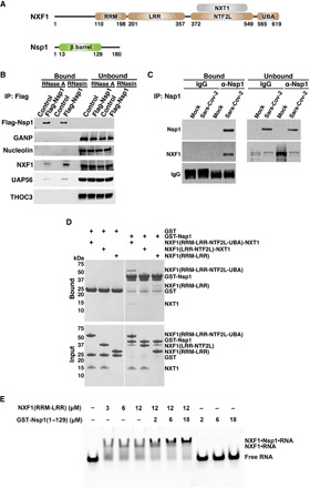
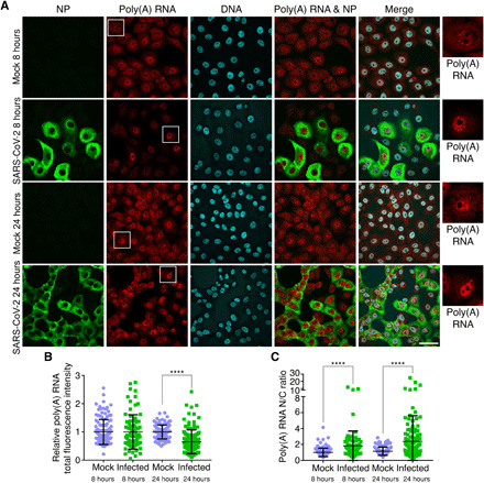
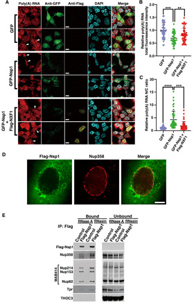
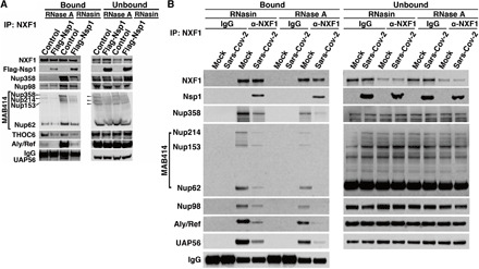
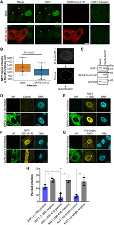
The ongoing unprecedented severe acute respiratory syndrome caused by the SARS-CoV-2 outbreak worldwide has highlighted the need for understanding viral-host interactions involved in mechanisms of virulence. Here, we show that the virulence factor Nsp1 protein of SARS-CoV-2 interacts with the host messenger RNA (mRNA) export receptor heterodimer NXF1-NXT1, which is responsible for nuclear export of cellular mRNAs. Nsp1 prevents proper binding of NXF1 to mRNA export adaptors and NXF1 docking at the nuclear pore complex. As a result, a significant number of cellular mRNAs are retained in the nucleus during infection. Increased levels of NXF1 rescues the Nsp1-mediated mRNA export block and inhibits SARS-CoV-2 infection. Thus, antagonizing the Nsp1 inhibitory function on mRNA export may represent a strategy to restoring proper antiviral host gene expression in infected cells.


Figures

Fig. 1
Fig. 1. Nsp1 protein of SARS-CoV-2 interacts with the mRNA nuclear export receptor NXF1.
(A) Schematic representation of NXF1-NXT1 and Nsp1. NXF1 contains an N-terminal RNA recognition motif (RRM), a leucine-rich repeat domain (LRR), a nuclear transport factor 2-like domain (NTF2L), and a ubiquitin-associated domain (UBA). RRM and LRR domains form an RNA binding module. NXT1 forms a heterodimer with NXF1 by binding to the NTF2L domain of NXF1. Nsp1 contains a β barrel domain. (B) 293T cells were transfected with 3xFlag-Nsp1 for 24 hours and then subjected to fractionation into nuclear and cytoplasmic fractions. See fig. S1 for fractionation controls. Nuclear lysates were subjected to immunoprecipitation (IP), followed by Western blot analysis to detect the indicated proteins. n = 3. (C) 293T cells expressing the SARS-CoV-2 receptor ACE2 protein were infected with SARS-CoV-2 at an MOI of 1 for 24 hours. Nsp1 was specifically immunoprecipitated from the cell lysates, and NXF1 interaction was detected by Western blot analysis. n = 3. (D) In vitro GST pull-down assays using the depicted purified recombinant proteins show that Nsp1 directly binds to NXF1. n = 3. (E) NXF1 associates with RNA in the presence of Nsp1. An electrophoretic mobility assay was carried out with a fluorescently labeled poly(U) 15-mer RNA and purified recombinant NXF1 (RRM-LRR) and/or Nsp1 (1-129) as indicated. n = 3.
Fig. 2
Fig. 2. SARS-CoV-2 inhibits poly(A) RNA nuclear export.
(A) Poly(A) RNA was assessed by RNA-FISH in Vero cells mock-infected or infected with SARS-CoV-2 at an MOI of 2 for 8 and 24 hours. Infected cells were detected with antibody against the viral NP protein. Boxed regions are enlarged and shown on the right. Scale bar, 50 μm. (B and C) Fluorescence intensities of total cellular poly(A) RNA in individual cells (B) or the N/C ratio of poly(A) RNA signals in individual cells (C) is shown for the various depicted conditions. Data represent three independent experiments. Mock, 8 hours, n = 126 cells; infected, 8 hours, n = 80 cells; mock, 24 hours, n = 134 cells; and infected, 24 hours, n = 133 cells. ****P < 0.0001.
Fig. 3
Fig. 3. NXF1 expression reverts Nsp1-mediated mRNA export block and reduction in mRNA levels.
(A) 293T cells were transfected with plasmids encoding GFP, GFP-Nsp1, or GFP-Nsp1 and Flag-NXF1 and then analyzed by RNA-FISH to detect poly(A) RNA and immunofluorescence to detect GFP, GFP-Nsp1, and Flag-NXF1. White arrowheads show examples for each condition. Scale bars, 10 μm. (B) Fluorescence intensity of total poly(A) RNA in individual cells is shown in the depicted conditions. (C) Fluorescence intensity of poly(A) in the nucleus and cytoplasm was determined, and the ratios of the nuclear-to-cytoplasmic signals for individual cells are shown for the indicated experimental condition. n = 3; ****P < 0.0001, ***P < 0.001, and **P < 0.01. (D) SK-N-SH cells were transfected with 3xFlag-Nsp1, and immunofluorescence microscopy was performed to detect Nsp1 and endogenous Nup358. n = 3. Scale bar, 5 μm. (E) Immunoprecipitation of Nsp1 followed by Western blot analysis shows Nsp1 interaction with certain nucleoporins, which is partially dependent on RNA.
Fig. 4
Fig. 4. Nsp1 prevents proper interaction of NXF1 with the mRNA export machinery.
(A) Western blot analysis of proteins immunoprecipitated with NXF1 in the presence or absence of Nsp1 or RNA reveals that the interaction of NXF1 with key mRNA export adaptors and certain Nups is inhibited by Nsp1. (B) 293T cells expressing the SARS-CoV-2 receptor ACE2 protein were infected with SARS-CoV-2 at an MOI of 1 for 24 hours. NXF1 was specifically immunoprecipitated from the cell lysates and followed by Western blot analysis to detect nucleoporins and mRNA export factors. Association of NXF1 with constituents of the mRNA export machinery is inhibited during SARS-CoV-2 infection.
Fig. 5
Fig. 5. NXF1 localization at the NPC is reduced upon SARS-CoV-2 infection and increased NXF1 level prevents infection.
(A) Vero E6 cells were infected with SARS-CoV-2 at an MOI of 1 for 48 hours. Fixed cells were treated with digitonin (1 μg/ml) to permeabilize the plasma membrane and examined by immunofluorescence microscopy to detect NXF1 and the viral NP protein. Scale bar, 10 μm. (B) The regions of the nuclear periphery were manually selected from equatorial sections of the nuclei in the z-stack images as indicated on the right, and the signal intensities along the NE were quantified (n = 22 for mock and n = 19 for SARS-CoV-2). Individual measurements (black dots) and the summarized box and whisker plots were overlaid (left). The difference in the NXF1 intensities at the NE between the mock and the SARS-CoV-2 infection samples was tested using Student’s t test. (C) The whole-cell extracts from Vero cells mock or SARS-CoV-2 infected were analyzed by Western blotting using anti-NXF1, SARS-CoV-2 NP, and GAPDH antibodies at 48 hours postinfection. The positions of molecular mass makers are indicated. (D to G) Vero E6 cells were transfected for 24 hours with plasmids encoding full-length Flag-tagged NXF1 or the indicated mutants of NXF1 and then infected with SARS-CoV-2 at an MOI of 1 for 24 hours. Cells were examined by immunofluorescence (IF) microscopy to detect viral NP protein and Flag-tagged NXF1. Scale bars, 10 μm. (H) Percentage of infected cells (NP+) was scored for each condition. Bars represent averages of three separate experiments, and dots represent the average of each of the three independent experiments. NXF1 (1–200), n = 831 cells; NXF1 (201–619), n = 1146 cells; and NXF1 (full-length), n = 1267 cells. **P < 0.01 and *P < 0.05.

Comment in

References

    1. Ayllon J., García-Sastre A., The NS1 protein: A multitasking virulence factor. Curr. Top. Microbiol. Immunol. 386, 73–107 (2015). - PubMed
    1. Satterly N., Tsai P.-L., van Deursen J., Nussenzveig D. R., Wang Y., Faria P. A., Levay A., Levy D. E., Fontoura B. M. A., Influenza virus targets the mRNA export machinery and the nuclear pore complex. Proc. Natl. Acad. Sci. U.S.A. 104, 1853–1858 (2007). - PMC - PubMed
    1. Zhang K., Xie Y., Muñoz-Moreno R., Wang J., Zhang L., Esparza M., García-Sastre A., Fontoura B. M. A., Ren Y., Structural basis for influenza virus NS1 protein block of mRNA nuclear export. Nat. Microbiol. 4, 1671–1679 (2019). - PMC - PubMed
    1. Zhang L., Das P., Schmolke M., Manicassamy B., Wang Y., Deng X., Cai L., Tu B. P., Forst C. V., Roth M. G., Levy D. E., García-Sastre A., de Brabander J., Phillips M. A., Fontoura B. M. A., Inhibition of pyrimidine synthesis reverses viral virulence factor-mediated block of mRNA nuclear export. J. Cell Biol. 196, 315–326 (2012). - PMC - PubMed
    1. Heath C. G., Viphakone N., Wilson S. A., The role of TREX in gene expression and disease. Biochem. J. 473, 2911–2935 (2016). - PMC - PubMed

Publication types

MeSH terms