Case Reports
. 2021 Feb 5;14(2):e239256.
doi: 10.1136/bcr-2020-239256.

Endovascular treatment of a sacral dural arteriovenous fistula

Affiliations
Case Reports

Endovascular treatment of a sacral dural arteriovenous fistula

Mokshal H Porwal et al. BMJ Case Rep. .

Abstract

Spinal dural arteriovenous fistula (SDAVF) is a rare pathological communication between arterial and venous vessels within the spinal dural sheath. Clinical presentation includes progressive spinal cord symptoms including gait difficulty, sensory disturbances, changes in bowel or bladder function, and sexual dysfunction. These fistulas are most often present in the thoracolumbar region. Diagnoses of SDVAFs are commonly missed, possibly due to the low index of suspicion, non-specific symptoms and challenging imaging. In this case report, we describe a rare presentation of a sacral SDAVF which was detected by collective efforts between endovascular neurosurgery and interventional radiology. We outline the diagnostic and imaging challenges we faced to discover the fistula. In particular, mechanical pump injection instead of hand injection during angiography was required to reveal the fistula. Following identification, the fistula was successfully treated endovascularly by using onyx (ethylene vinyl alcohol glue), a less invasive alternative to surgical intervention.

Keywords: interventional radiology; neurosurgery.


Conflict of interest statement

Competing interests: None declared.

Figures

Figure 1
Figure 1
Lateral thoracic MRI (A) and lateral lumbar MRI (B). Thoracic MRI shows dilated pial/subdural vessels and spinal cord oedema within the lower thoracic and conus region, noted by the green arrows (A). Lumbar MRI was consistent with no further notable characteristics (B). This is a classical appearance for a dural arteriovenous fistula.
Figure 2
Figure 2
Sagittal thoracic MRA. Thoracic MRA shows a prominent tortuous vessel along the left T8–T9 level suspicious for a spinal dural arteriovenous fistula (red arrows).
Figure 3
Figure 3
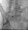
Anteroposterior projection with hand injection (A) and power injector (B) of left internal iliac artery angiogram. Note that the radicular draining vein is now visualised following power injection angiogram and glucagon was used to optimise image quality by reducing peristaltic bowel shadows in (B).
Figure 4
Figure 4
Note the venous pouch (red arrow) that exists within the fistulous epidural network prior to the radicular vein exiting. Microcatheter injection of feeding artery arising from left lateral sacral artery demonstrating a radiculomedullary vein running along nerve root as it exits the fistula. The radicular vein drains superiorly and then enters the congested medullary venous plexus (yellow arrow).
Figure 5
Figure 5
Final result of endovascular obliteration using onyx glue, demonstrating the cast within the venous pouch and draining radicular vein.

References

    1. Thron A Spinale durale arteriovenöse Fisteln [Spinal dural arteriovenous fistulas]. Radiologe 2001;41:955–60. - PubMed
    1. Jellema K, Canta LR, Tijssen CC, et al. . Spinal dural arteriovenous fistulas: clinical features in 80 patients. J Neurol Neurosurg Psychiatry 2003;74:1438–40. 10.1136/jnnp.74.10.1438 - DOI - PMC - PubMed
    1. Marcus J, Schwarz J, Singh IP, et al. . Spinal dural arteriovenous fistulas: a review. Curr Atheroscler Rep 2013;15:335. 10.1007/s11883-013-0335-7 - DOI - PubMed
    1. Ren Y, Liu H, Chen T-Y, et al. . Successful management of sacral dural arteriovenous fistulas: a case series and literature review. World Neurosurg 2019;126:164–70. 10.1016/j.wneu.2019.02.129 - DOI - PubMed
    1. Chen CJ, Chen CM, Lin TK. Enhanced cervical MRI in identifying intracranial dural arteriovenous fistulae with spinal perimedullary venous drainage. Neuroradiology 1998;40:393–7. 10.1007/s002340050609 - DOI - PubMed

Publication types

MeSH terms