. 2021 Apr;9(4):441-453.
doi: 10.1158/2326-6066.CIR-20-0451. Epub 2021 Feb 5.

Modifications to the Framework Regions Eliminate Chimeric Antigen Receptor Tonic Signaling

Affiliations

Modifications to the Framework Regions Eliminate Chimeric Antigen Receptor Tonic Signaling

Elisa Landoni et al. Cancer Immunol Res. 2021 Apr.

Abstract

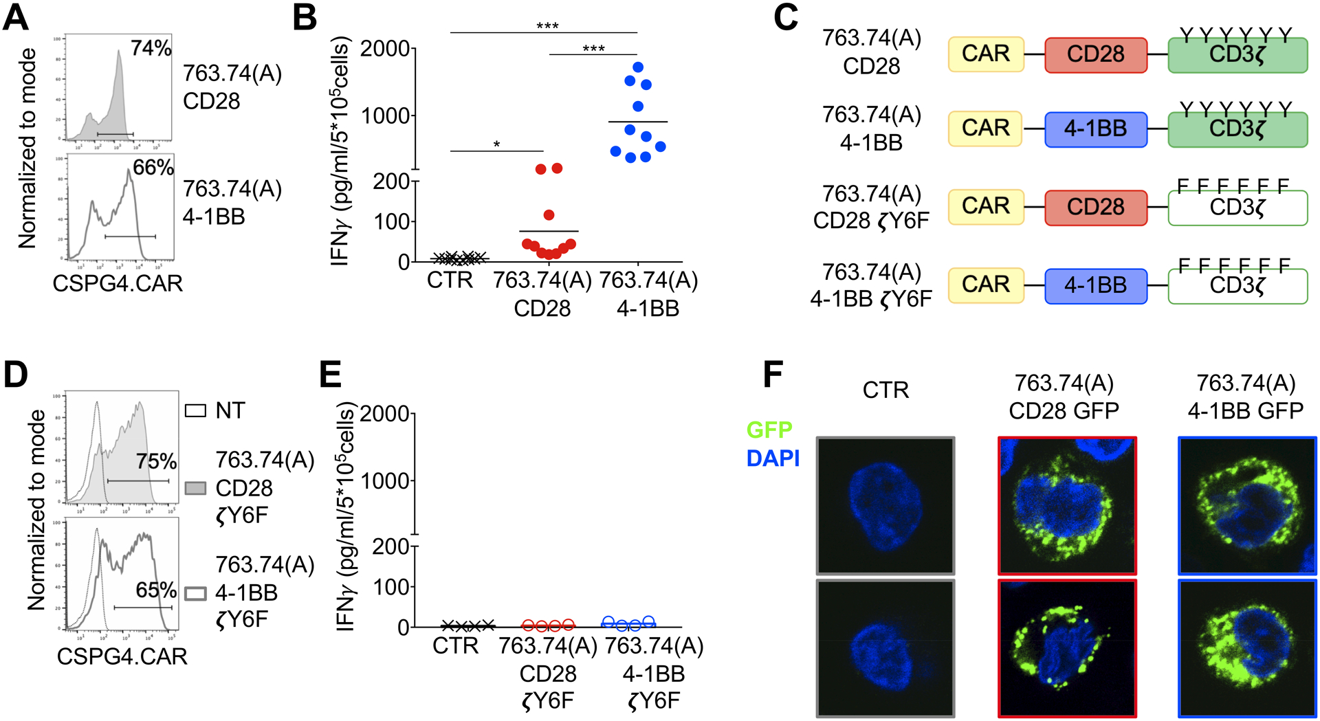
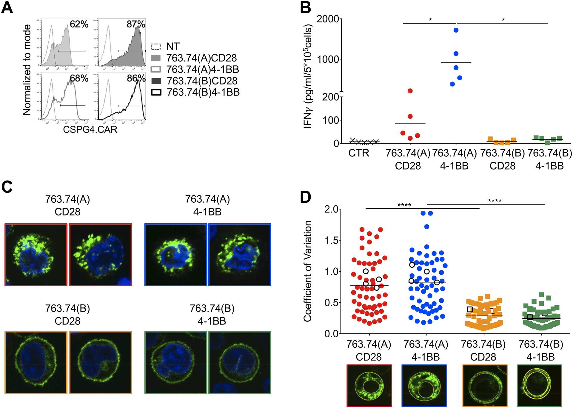
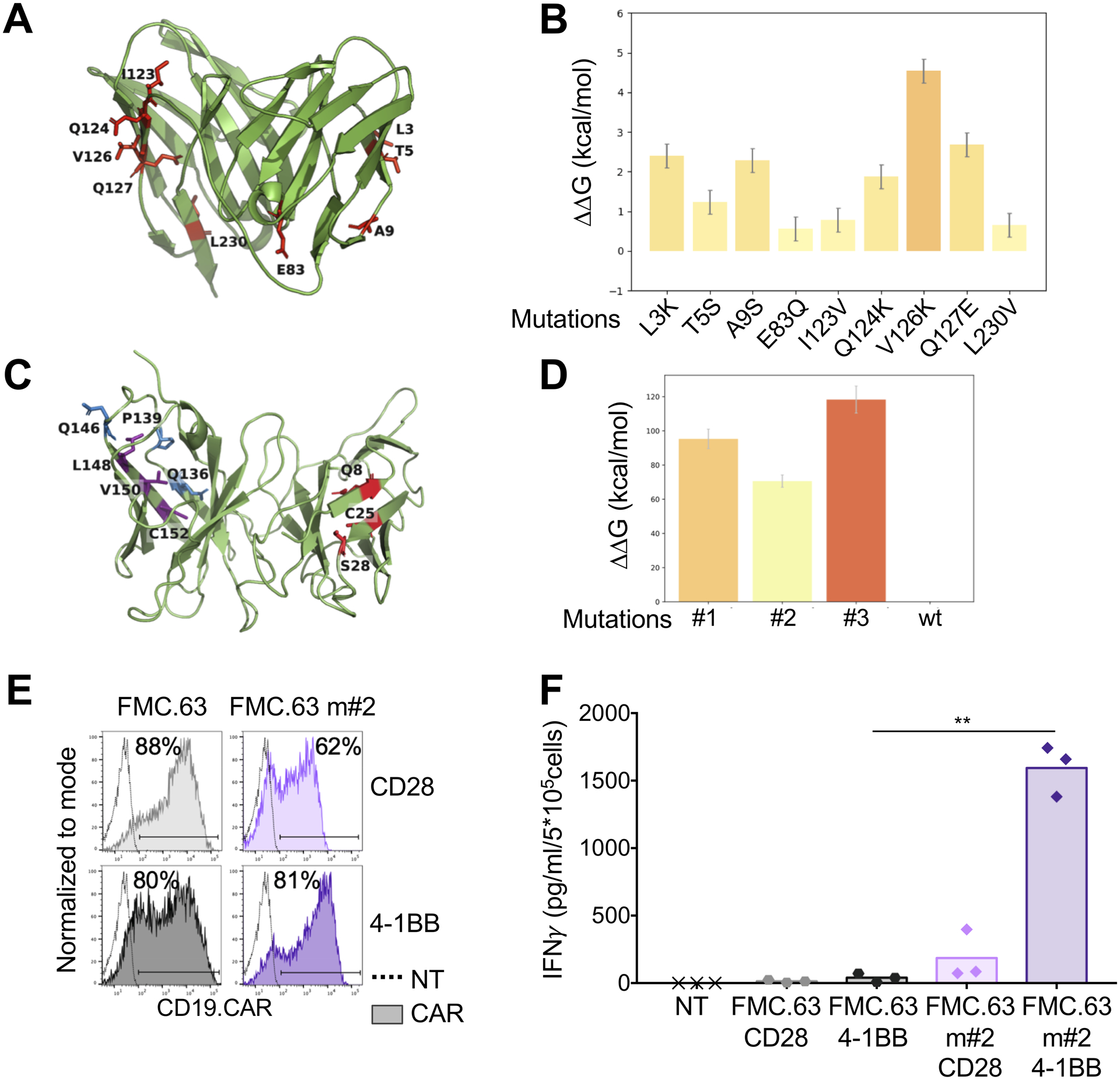
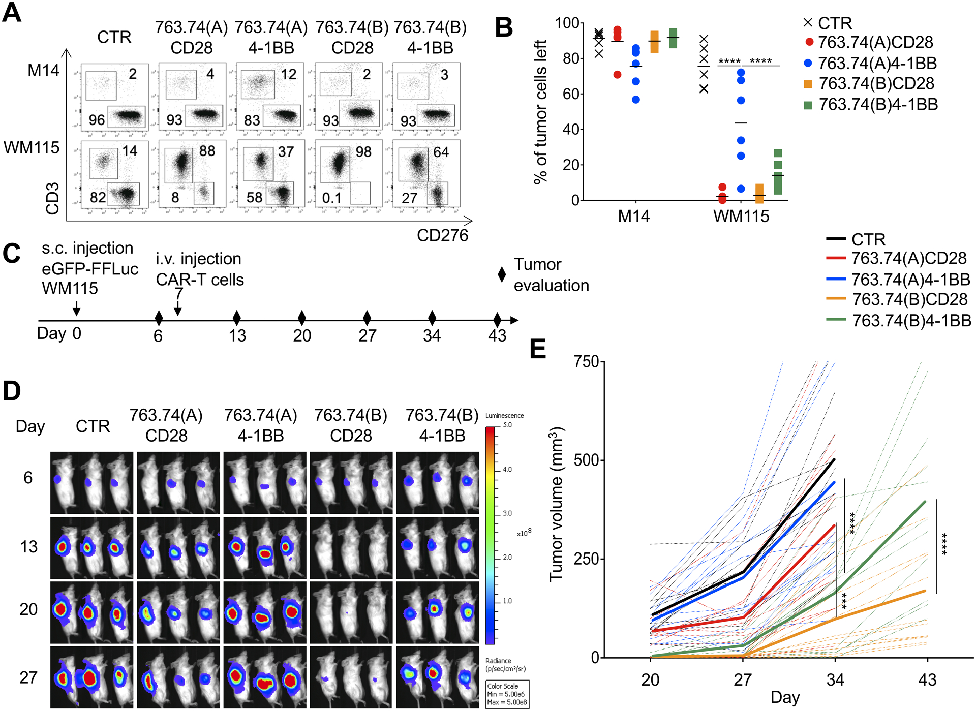
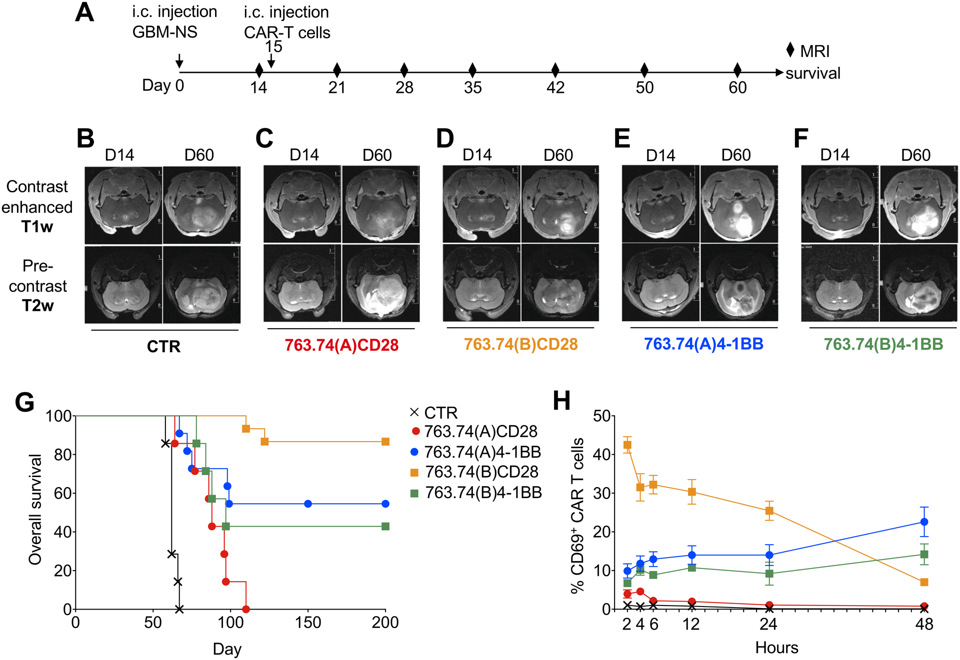
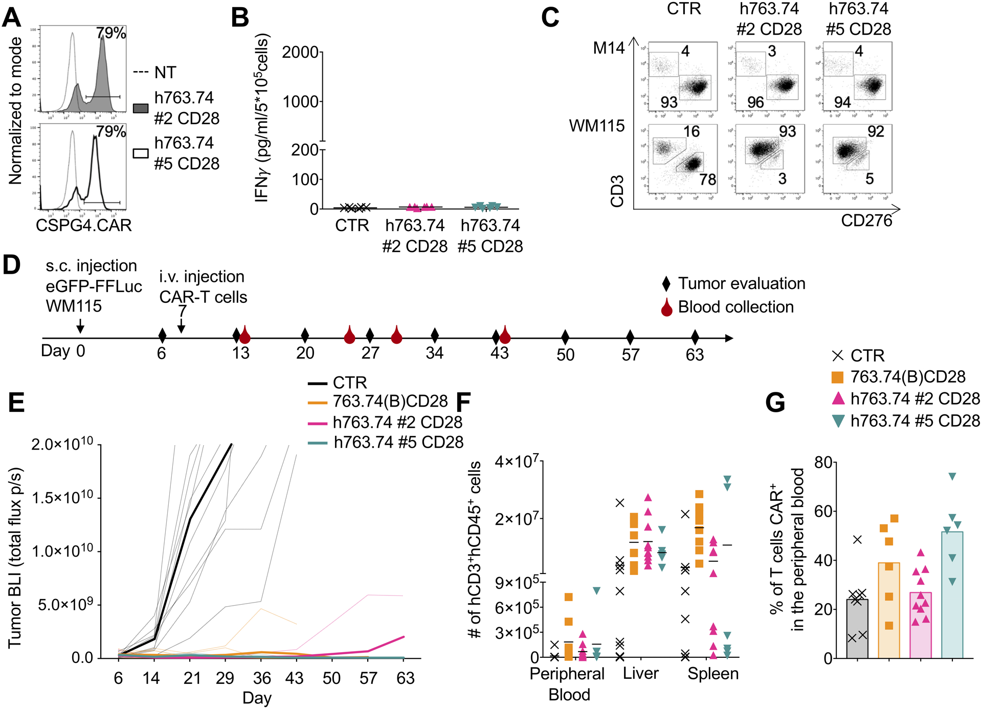
Chimeric antigen receptor (CAR) tonic signaling, defined as spontaneous activation and release of proinflammatory cytokines by CAR-T cells, is considered a negative attribute because it leads to impaired antitumor effects. Here, we report that CAR tonic signaling is caused by the intrinsic instability of the mAb single-chain variable fragment (scFv) to promote self-aggregation and signaling via the CD3ζ chain incorporated into the CAR construct. This phenomenon was detected in a CAR encoding either CD28 or 4-1BB costimulatory endodomains. Instability of the scFv was caused by specific amino acids within the framework regions (FWR) that can be identified by computational modeling. Substitutions of the amino acids causing instability, or humanization of the FWRs, corrected tonic signaling of the CAR, without modifying antigen specificity, and enhanced the antitumor effects of CAR-T cells. Overall, we demonstrated that tonic signaling of CAR-T cells is determined by the molecular instability of the scFv and that computational analyses of the scFv can be implemented to correct the scFv instability in CAR-T cells with either CD28 or 4-1BB costimulation.


Conflict of interest statement

Disclosure of Potential Conflicts of Interest

Drs Dotti, Savoldo and Ferrone hold patents in the field of T cell engineering and have sponsor research agreements with Bluebird Bio and Bellicum Pharmaceutical. Dr. Dotti serves in the scientific advisory board of Bellicum Pharmaceutical and Catamaran.

Figures

Figure 1.
Figure 1.. Tonic signaling of CAR-T cells expressing the CAR encoding scFv 763.74(A).
A. Representative flow cytometry plots showing CAR expression in T cells assessed at day 8 of culture. 763.74(A) CD28 and 763.74(A) 4–1BB indicate the specific CAR expressed in T cells. CAR expression was assessed using an anti-idyotipic antibody followed by the staining with a secondary rat anti-mouse antibody. B. Quantification of IFNγ released into supernatants by T cells expressing control CAR (CTR), 763.74(A) CD28, or 763.74(A) 4–1BB CAR without CAR-specific activation after 24 hours (n=10, mean shown). *P=0.0184; ***P=0.0003 (CTR vs. 763.74(A) CD28); ***P=0.0004 (763.74(A) CD28 vs. 763.74(A) 4–1BB), paired t-test. C. Schema of the 763.74(A) CAR constructs encoding either CD3ζ wild-type or CD3ζ in which all tyrosine amino acids of ITAMs have been mutated. D. Representative flow cytometry plots showing CAR expression in T cells assessed at day 8 of culture. 763.74(A) CD28 ζY6F and 763.74(A)4–1BB ζY6F indicate the specific CAR expressed in T cells. CAR expression was assessed using an anti-idyotipic antibody followed by the staining with a secondary rat anti-mouse antibody. Non-transduced (NT) T cells were a negative control. E. Quantification of IFNγ released into supernatants by T cells expressing control CAR (CTR), 763.74(A) CD28ζY6F or 763.74(A) 4–1BBζY6F CAR without CAR-specific activation after 24 hours (n=4, mean shown). F. Representative confocal microscopy imaging showing GFP aggregation in T cells expressing GFP-tagged CARs (green) in which the CARs are obtained using the scFv 763.74(A) and either CD28 or 4–1BB endodomains. CTR indicates control T cells. Blue staining indicates the DAPI. Shown are representative cell of a single field (Magnification 63X).
Figure 2.
Figure 2.. Amino acid substitutions in the FWRs of the scFv 763.74(A) reverse the tonic signaling in CAR-T cells.
A. Representative flow cytometry plots showing CAR expression in T cells engineered with the scFv 763.74(A) and scFv 763.74(B) CARs encoding either CD28 or 4–1BB endodomains. CAR expression was assessed using an anti-idyotipic antibody followed by the staining with a secondary rat anti-mouse antibody. Non-transduced (NT) T cells were a negative control. B. Quantification of IFNγ released into supernatants by T cells expressing the different CARs without CAR-specific activation after 24 hours. CTR: control CAR-T cells (n=5, mean shown). *P=0.0154 763.74(A) CD28 vs. 4–1BB; *P=0.194 763.74(A) vs. (B)4–1BB, paired t test. C. Representative confocal microscopy imaging showing GFP aggregation in T cells expressing GFP-tagged CARs in which the CARs were obtained using either the scFv 763.74(A) or scFv 763.74(B) and either CD28 or 4–1BB endodomains. Blue staining indicates the DAPI. Shown are representative cells of a single field (Magnification 63X). D. Quantification of the relative variability of GFP-tagged CAR expression using the Coefficient of Variation calculated as standard deviation/average pixel MFI. The standard deviation and average pixel MFI were calculated on the GFP+ areas. White symbols represent the cells shown in Fig. 1F and Fig. 2C (n=60, mean shown). ****P<0.0001, unpaired t-test.
Figure 3.
Figure 3.. Amino acid substitutions in the FWRs of the scFv 763.74(A) destabilize the scFv.
A. Structural conformation of the scFv 763.74(B) generated through computational modeling. Protein is shown in cartoon representation and FWR mutations in stick representation. B. Chosen amino acid mutations evaluated for their influence on scFv 763.74(B) stability (n=50, mean and SD shown). C. Structural conformation of the scFv FMC.63 generated through computational modeling. Protein is shown in cartoon representation and FWR mutations in stick representation. D. Chosen amino acid mutations (Q8W|C25R|S28P, Q136W|P139L|Q146P, and L148Y|V150W|C152W) evaluated for their influence on the scFv FMC.63 stability (n=50, mean and SD shown). E. Representative flow cytometry plots showing CAR expression in T cells engineered with the scFv FMC.63 CARs and the destabilized scFv FMC.63 m#2 CARs encoding either CD28 or 4–1BB endodomains. CAR expression was assessed using an anti-idyotipic antibody followed by the staining with a secondary rat anti-mouse antibody. F. Quantification of IFNγ released into supernatants by T cells expressing the different CARs without CAR-specific activation after 24 hours (n=3, mean shown). **P=0.0047, paired t test.
Figure 4.
Figure 4.. T cells expressing the 763.74(B) CAR with CD28 are superior in mediating tumor elimination in a melanoma tumor model.
A Representative flow plots and B. summary of the quantification of residual tumor cells of experiments in which control T cells (CTR) and T cells engineered with the scFv 763.74(A) and scFv 763.74(B) CARs encoding either CD28 or 4–1BB endodomains. CAR-T cells were cocultured with melanoma cell lines (E:T=1:5) for 5 days and then collected and stained with anti-CD3 and anti-CD276 (B7-H3) to identify T cells and melanoma cells, respectively, by flow cytometry. Data are presented as mean±SD, n=6. ****P<0.0001, two-way ANOVA with Bonferroni’s correction. C. Experimental schema of the melanoma xenograft model. eGFP-FFLuc WM115 (5 × 10⁵ cells) were injected s.c., and 7 days later, mice were injected i.v. with control T cells (CTR) or T cells engineered with the scFv 763.74(A) and scFv 763.74(B) CARs encoding either CD28 or 4–1BB endodomains (5×10⁶ cells). D. Representative tumor bioluminescence (BLI)(color scale: min=5 × 10⁶; max=5 × 10⁸) in mice treated as illustrated in (C). E. Tumor volume in mice engrafted in (D). Dotted lines represent individual mice, and bolded solid lines represent the mean for the group. Summary of 4 independent experiments (n=12 for each condition). ***P=0.00012; ****P<0.0001, two-way ANOVA with Bonferroni’s correction.
Figure 5.
Figure 5.. T cells expressing the 763.74(B) CAR with CD28 have rapid antitumor activity in a glioblastoma tumor model.
A. Experimental schema of the GBM xenograft model. GBM-NS (1×10⁵ cells) were injected into the caudate nucleus, and 15 days later, mice were injected intratumorally with control T cells (CTR) or T cells engineered with the scFv 763.74(A) and scFv 763.74(B) CARs encoding either CD28 or 4–1BB endodomains (2×10⁶ cells). Tumor growth was monitored with MRI. B-F. Representative MRI performed with T1-weighted images (T1-wi) with contrast medium injection and T2-weighted images (T2-wi) showing the pattern of tumor progression and infiltration in mice treated as in (A). G. Kaplan-Meier survival curves of mice treated as in (A). n=7 mice/group for CTR, 763.74(A)CD28, 763.74(B)4–1BB. n=15 mice/group for 763.74(B) CD28 and 763.74(A) 4–1BB. Overall survival statistical analysis was performed using the Mantel-Cox log rank test. H. Time course of CD69 expression in CAR-T cells isolated from the tumor at the indicated time points after intratumor delivery of T cells. Data are presented as mean±SD, n=2.
Figure 6.
Figure 6.. Humanization of the FWRs of the scFv 763.74(A) abrogates CAR tonic signaling without affecting the anti-tumor activity.
A. Representative flow cytometry plots showing CAR expression in T cells engineered with h763.74 #2 and h763.74 #5 CARs encoding CD28 (h763.74 #2 CD28 and h763.74 #5 CD28) as assessed at day 8 of culture. CAR expression was assessed using a biotinylated antibody recognizing the human FWRs followed by the staining with streptavidin protein. Non-transduced (NT) T cells were a negative control. B. Quantification of IFNγ in supernatants of control (CTR), h763.74 #2 CD28, and h763.74 #5 CD28 CAR-T cells collected after 24 hours. Data are presented as mean±SD, n=6. C. Representative flow plots of coculture experiments in which control (CTR) or T cells expressing either h763.74 #2 CD28 or h763.74 #5 CD28 CARs were plated with melanoma cell lines (E:T=1:5) for 5 days. Cells were then collected and stained with the anti-CD3 and anti-CD276 to identify T cells and melanoma cells, respectively, by flow cytometry. D. Experimental schema of the melanoma xenograft model. eGFP-FFLuc WM115 (5×10⁵ cells) were injected s.c., and 7 days later, mice were injected i.v. with control T cells (CTR) or T cells engineered with 763.74(B) CD28 CAR, h763.74 #2 CD28, or h763.74 #5 CD28 CARs (5×10⁶ cells). E. Tumor BLI kinetics of mice treated according to scheme (D). Dotted lines represent individual mice, and bolded solid lines represent the mean for the group. Summary of 2 independent experiments (n=10 for each group). F. Quantification of human CD3+CD45+ cells in the peripheral blood, liver, and spleen at the time of euthanasia of tumor-bearing mice treated as in (D). Data are presented as mean±SD, n=6. G. Percentage of CAR-T cells in the peripheral blood, gated on human CD3+CD45+ cells, at sacrifice in tumor-bearing mice treated as in (D). Data are presented as mean±SD, n=6.

References

    1. Eshhar Z, Waks T, Gross G, and Schindler DG 1993. Specific activation and targeting of cytotoxic lymphocytes through chimeric single chains consisting of antibody-binding domains and the gamma or zeta subunits of the immunoglobulin and T-cell receptors. Proc. Natl. Acad. Sci. U. S. A 90:720–724. - PMC - PubMed
    1. Finney HM, Lawson AD, Bebbington CR, and Weir AN 1998. Chimeric receptors providing both primary and costimulatory signaling in T cells from a single gene product. J. Immunol 161:2791–2797. - PubMed
    1. Imai C, Mihara K, Andreansky M, Nicholson IC, Pui CH, Geiger TL, and Campana D 2004. Chimeric receptors with 4–1BB signaling capacity provoke potent cytotoxicity against acute lymphoblastic leukemia. Leukemia 18:676–684. - PubMed
    1. Maude SL, Frey N, Shaw PA, Aplenc R, Barrett DM, Bunin NJ, Chew A, Gonzalez VE, Zheng Z, Lacey SF et al. 2014. Chimeric antigen receptor T cells for sustained remissions in leukemia. N. Engl. J. Med 371:1507–1517. - PMC - PubMed
    1. Dotti G, Gottschalk S, Savoldo B, and Brenner MK 2014. Design and development of therapies using chimeric antigen receptor-expressing T cells. Immunol. Rev 257:107–126. - PMC - PubMed

Publication types

MeSH terms