. 2021 Feb 9;118(6):e2011038118.
doi: 10.1073/pnas.2011038118.

Diel transcriptional oscillations of light-sensitive regulatory elements in open-ocean eukaryotic plankton communities

Affiliations

Diel transcriptional oscillations of light-sensitive regulatory elements in open-ocean eukaryotic plankton communities

Sacha N Coesel et al. Proc Natl Acad Sci U S A. .

Abstract

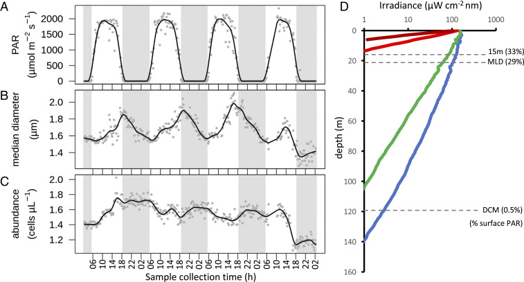
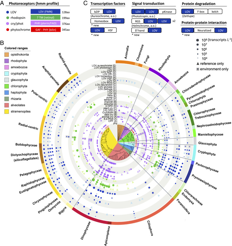
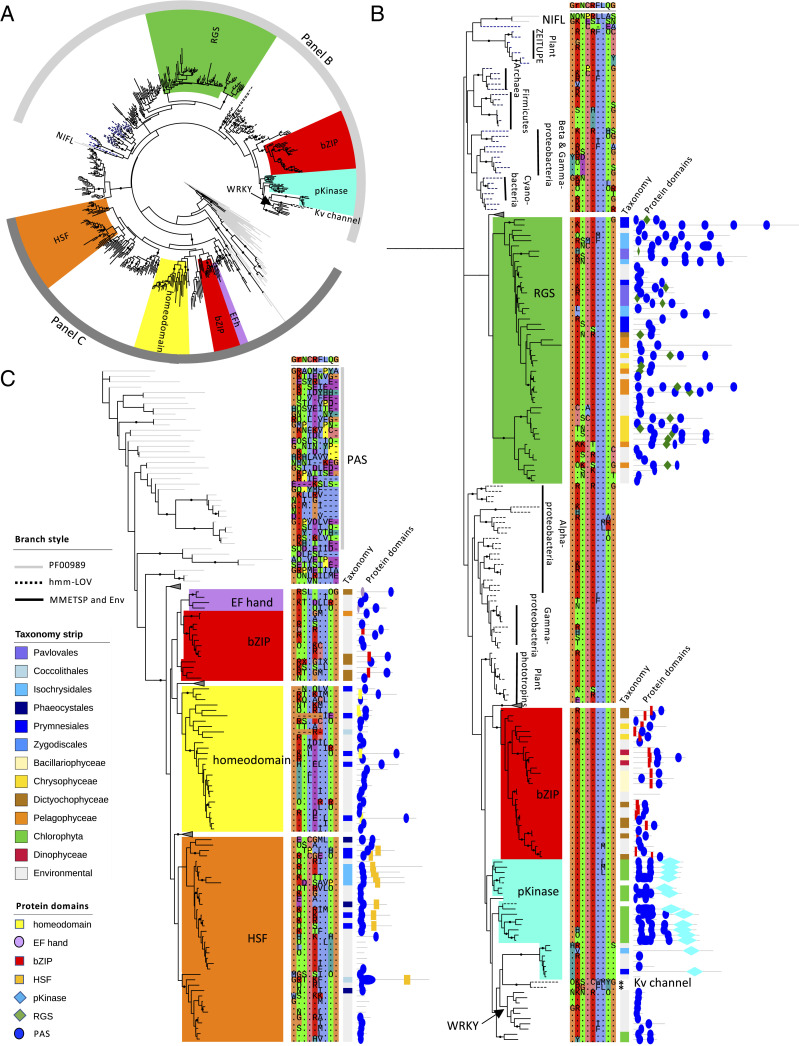
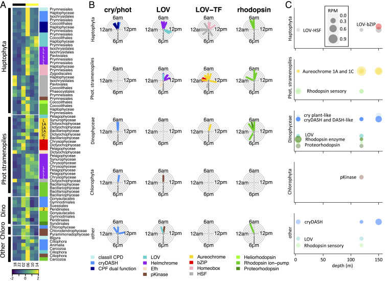
The 24-h cycle of light and darkness governs daily rhythms of complex behaviors across all domains of life. Intracellular photoreceptors sense specific wavelengths of light that can reset the internal circadian clock and/or elicit distinct phenotypic responses. In the surface ocean, microbial communities additionally modulate nonrhythmic changes in light quality and quantity as they are mixed to different depths. Here, we show that eukaryotic plankton in the North Pacific Subtropical Gyre transcribe genes encoding light-sensitive proteins that may serve as light-activated transcription factors, elicit light-driven electrical/chemical cascades, or initiate secondary messenger-signaling cascades. Overall, the protistan community relies on blue light-sensitive photoreceptors of the cryptochrome/photolyase family, and proteins containing the Light-Oxygen-Voltage (LOV) domain. The greatest diversification occurred within Haptophyta and photosynthetic stramenopiles where the LOV domain was combined with different DNA-binding domains and secondary signal-transduction motifs. Flagellated protists utilize green-light sensory rhodopsins and blue-light helmchromes, potentially underlying phototactic/photophobic and other behaviors toward specific wavelengths of light. Photoreceptors such as phytochromes appear to play minor roles in the North Pacific Subtropical Gyre. Transcript abundance of environmental light-sensitive protein-encoding genes that display diel patterns are found to primarily peak at dawn. The exceptions are the LOV-domain transcription factors with peaks in transcript abundances at different times and putative phototaxis photoreceptors transcribed throughout the day. Together, these data illustrate the diversity of light-sensitive proteins that may allow disparate groups of protists to respond to light and potentially synchronize patterns of growth, division, and mortality within the dynamic ocean environment.

Keywords: diel cycles; metatranscriptomics; microbial eukaryotes; oligotrophic gyre; photoreceptors.


Conflict of interest statement

The authors declare no competing interest.

Figures

Fig. 1.
Fig. 1.
Characteristics of the sampling site near Station ALOHA (July 26 to 30, 2015). (AC) Photosynthetically active light (PAR) intensity at the ocean surface (A), median cell diameter of eukaryotic phytoplankton less than 5 μm in diameter (B), and abundance of eukaryotic phytoplankton less than 5 μm in diameter (C). Points indicate measurements, and solid lines represent smoothed data (spline of order 3). Collection times of metatranscriptome samples are indicated. (D) Depth profile of available irradiance at wavelengths of 430 to 480nm (blue line), 500 to 560 nm (green line), 650 to 680 nm (light red line), and 700 to 740 nm (dark red line) were measured at noon on July 30, 2015. Dashed lines indicate the 15-m sampling depth, the 21-m mixed-layer depth (MLD), and the 119-m DCM; the percentage of PAR as compared to the surface PAR is indicated for these depths.
Fig. 2.
Fig. 2.
Abundance and taxonomic distribution of environmental photoreceptor and other light-sensitive protein-encoding transcripts. (A) Schematic representation of the hmm-profiles used to identify environmental photoreceptor and other light-sensitive protein-encoding transcripts. The respective chromophores are indicated with parentheses: flavin mononucleotide (FMN) for the LOV domain, retinal for the seven transmembrane (7 TM) helices of rhodopsin, pterin and flavin adenine dinucleotide (FAD) for the photolyase-homologous region (PHR) of the CPF, and bilin for the GAF and PHY domain constituting the photosensory part of phytochrome. The length of the hmm-profile is indicated in amino acids (aa). (B) Environmental transcript abundance of phytochrome (red), cryptochromes/photolyase (violet), rhodopsin (green), and those with LOV domains (blue) visualized on an 18S ribosomal RNA maximum-likelihood phylogenetic tree, representing 117 different eukaryotic orders relevant for the marine environment. The taxonomic phylum and class-level classifications are indicated by the colored ranges further annotated in SI Appendix, Fig. S6. Protein subtypes (clades A ∼ E) are derived from the respective reference trees (SI Appendix, Figs. S1–S4). Colored circles indicate transcripts detected in both the reference sequences and the environmental samples at the respective order level. The size of the circle corresponds to the mean environmental transcript concentrations (transcripts per liter) over the 4-d sampling period. Triangles indicate detection of transcripts in the reference sequences that are not detected in the environmental samples. Gray boxed circles indicate transcripts detected in the environmental samples only. (C) Schematic presentation of the domain structures of the LOV domain-containing sequences retrieved from our light-sensitive protein database of reference sequences. 6-4 Phot, (6-4) photolyase; animal cry, animal type I cryptochrome; bact cry, bacterial cryptochrome; channel, channel rhodopsin; CPF, (6-4) photolyase/cryptochrome dual-function proteins; CryDASH, cryptochrome-DASH; enzyme, enzyme rhodopsin; helio, heliorhodopsin; I-III CPD, type I to III CPD photolyase; II CPD, type II CPD photolyase; LOV (A ∼ E), clade-aggregated counts of LOV transcripts (SI Appendix, Fig. S3) that are not included in B; plant cry, plant cryptochrome (found only in Chlorophyta and Rhodophyta) and plant-like cryptochrome (all other taxonomies; SI Appendix, Fig. S3); phy, phytochrome; pump, proteorhodopsin and other ion-pump rhodopsins; sensory, sensory rhodopsin.
Fig. 3.
Fig. 3.
Phylogenetic and domain analysis of LOV-containing environmental contigs. (A) Maximum-likelihood tree (RAxML) of protist environmental LOV-domain contigs that are transcribed with a diel rhythmicity and their closest reference database-derived homologs (black edges). Also included are PAS-domain (PF00989; gray edges) and LOV-domain (hmm-LOV; dashed edges) reference sequences. Effector domains are indicated by the colored ranges. Bootstrap values of 80% and higher (100 iterations) are indicated with black circles. The gray bars indicate the upper and lower sections of the tree that are expanded in B and C, respectively. The arrow indicates the placement site of the WRKY-LOV sequences (SI Appendix, Fig. S9) by pplacer. (B and C) Expansion of the upper and lower tree sections, respectively. Shown are only the clades in which the LOV domain was found to be associated with an effector domain. Indicated from left to right: alignment of the light-sensitive motif GXNCRFLQG within the LOV domain (ClustalX colorscheme; dotted repeats), color strip indicating the taxonomies of the sequences retrieved from our light-sensitive protein database with the environmental-derived contigs in gray, and schematic representation of the protein sequences with the locations of the protein domain motifs indicated by the various shapes color-coded as the tree ranges and with the PAS/LOV domain in blue. The non–light-sensitive NIFL and Kv channel sequences are indicated. The asterisk indicates that the light-sensitive motifs of the Kv channel proteins contain two additional amino acid residues (see also SI Appendix, Fig. S8).
Fig. 4.
Fig. 4.
Diel and depth signatures of environmental photoreceptor and other light-sensitive protein-encoding transcripts. (A) Abundance (z score-normalized) of transcripts that displayed a 24-h diel rhythmicity (RAIN; P < 0.001), clustered at the phylogenetic order level for the three dominant eukaryotic phyla: Haptophyta, photosynthetic stramenopiles, and Dinophyceae. The color strip next to the heat map marks the different protein types. Subtypes are indicated for aureochrome 1A, 1C, and 2 and for aureochrome- and helmchrome-like (L). Taxonomies are indicated. (B) Modeled peak times (mFourfit) for each class of protein with a predicted period between 23 and 25 h are indicated in the radial plots. The length of the pointer corresponds to the mean transcript abundance in log scale, with the inner ring corresponding to 101 reads per liter and the outer ring corresponding to 107 reads per liter. The pointers are color-coded by protein type as in A. (C) The top 25 most abundant light-sensitive protein-encoding transcripts at three depths (5, 119, 150 m) in metatranscriptomic datasets collected at 1800 hours on day 4 of the cruise. The protein classes are color-coded as in A and also written within the charts. The size of the circles corresponds to the RPM values.

Similar articles

Cited by

References

    1. Roenneberg T., Merrow M., Circadian clocks - The fall and rise of physiology. Nat. Rev. Mol. Cell Biol. 6, 965–971 (2005). - PubMed
    1. Wijnen H., Young M. W., Interplay of circadian clocks and metabolic rhythms. Annu. Rev. Genet. 40, 409–448 (2006). - PubMed
    1. Noordally Z. B., Millar A. J., Clocks in algae. Biochemistry 54, 171–183 (2015). - PubMed
    1. Somers D. E., Phytochromes and cryptochromes in the entrainment of the Arabidopsis circadian clock. Science 282, 1488–1490 (1998). - PubMed
    1. Cashmore A. R., Cryptochromes: Enabling plants and animals to determine circadian time. Cell 114, 537–543 (2003). - PubMed

Publication types

MeSH terms

LinkOut - more resources