. 2021 Feb 5;12(1):828.
doi: 10.1038/s41467-021-21063-0.

Structural elements in the flexible tail of the co-chaperone p23 coordinate client binding and progression of the Hsp90 chaperone cycle

Affiliations

Structural elements in the flexible tail of the co-chaperone p23 coordinate client binding and progression of the Hsp90 chaperone cycle

Maximilian M Biebl et al. Nat Commun. .

Abstract

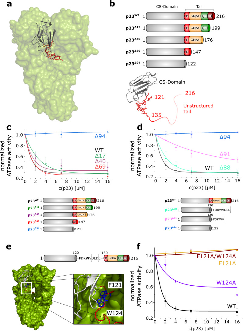
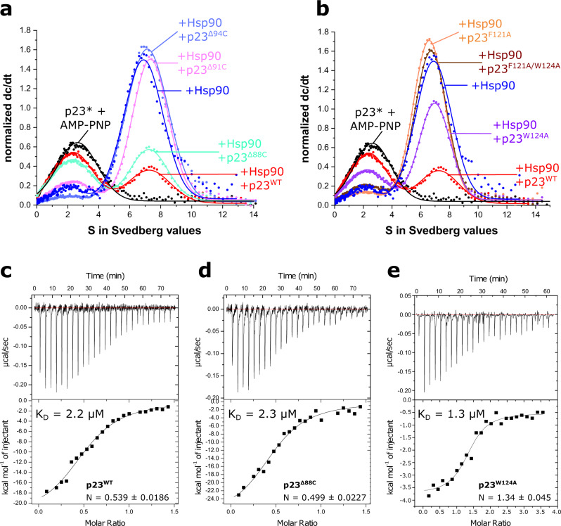
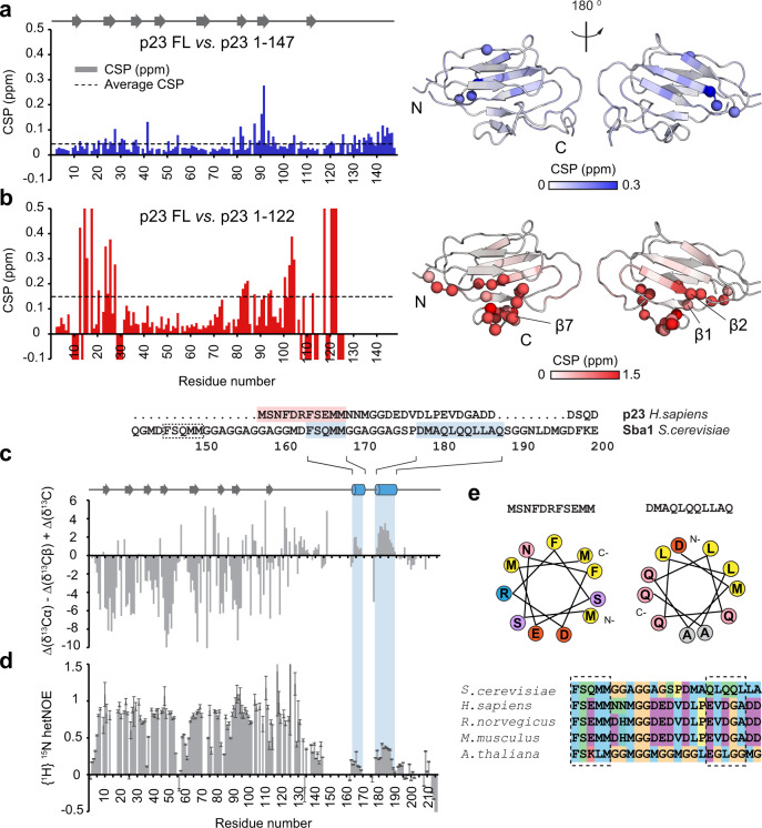
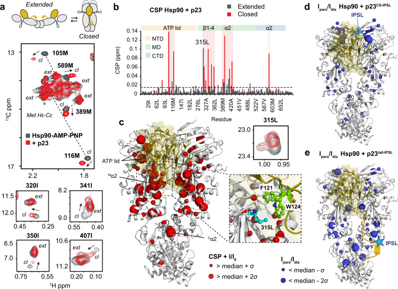
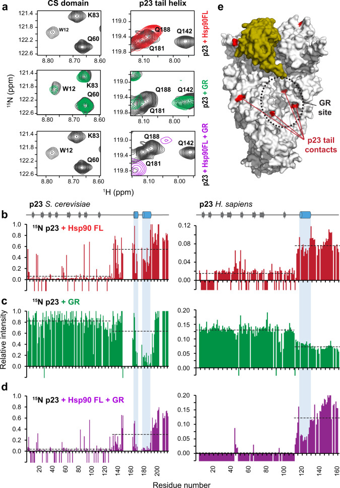
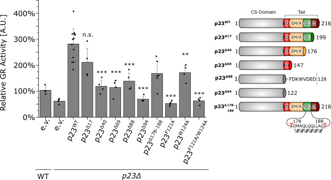
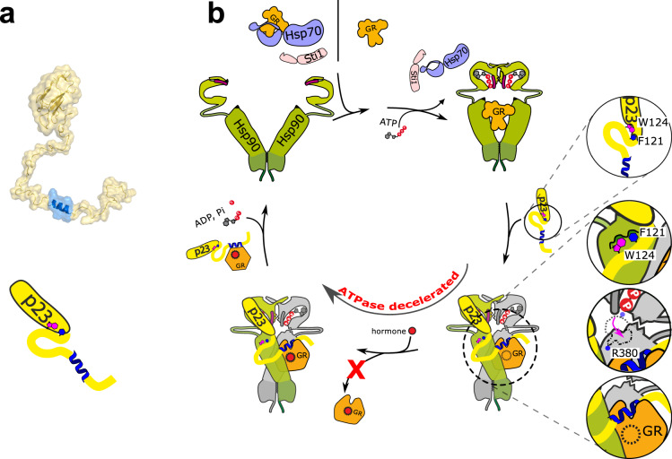
The co-chaperone p23 is a central part of the Hsp90 machinery. It stabilizes the closed conformation of Hsp90, inhibits its ATPase and is important for client maturation. Yet, how this is achieved has remained enigmatic. Here, we show that a tryptophan residue in the proximal region of the tail decelerates the ATPase by allosterically switching the conformation of the catalytic loop in Hsp90. We further show by NMR spectroscopy that the tail interacts with the Hsp90 client binding site via a conserved helix. This helical motif in the p23 tail also binds to the client protein glucocorticoid receptor (GR) in the free and Hsp90-bound form. In vivo experiments confirm the physiological importance of ATPase modulation and the role of the evolutionary conserved helical motif for GR activation in the cellular context.


Conflict of interest statement

The authors declare no competing interests.

Figures

Fig. 1
Fig. 1. p23-mediated inhibition of the Hsp90 ATPase activity depends on F121 and W124.
a The crystal structure of yeast p23 (Sba1) bound to yeast Hsp90 (Hsp82) is shown (PDB: 2CG9). The p23 CS domain is depicted in gray, the part of the p23 tail that was resolved in the crystal structure is shown in red. b The schematic domain architecture of p23 and the truncation mutants used in this study are shown. A close-up view of the p23 molecule is shown at the bottom (PDB: 2CG9). ED = negatively charged stretch; GM/A = GM/A-rich region; QL = QL-rich region; ED = second negatively charged stretch. cf All subfigures show ATPase assays in the presence of WT or mutant yeast p23 (Sba1). The ATPase activity of yeast Hsp90 (Hsp82) was determined in the presence of rising p23 concentrations. WT (black) is shown as a reference in all figures. c, d Effects of the deletion mutants p23Δ17, p23Δ40, p23Δ69 and p23Δ94 on the ATPase activity of Hsp90. Note that the same data set was used for p23WT and p23Δ94 in (c) and (d) as a reference. e Top: the p23 domain architecture is shown and the F121DKW124VD motif is highlighted. Bottom: crystal structure of p23 (gray cartoon) bound to the Hsp90 dimer (green surface) (PDB: 2CG9). The p23 CS domain is shown in gray, the tail region (up to aa 135) is depicted in red and the F121 and W124 residues are shown in blue. f Effect of the point mutants p23F121A, p23W124A and the double mutant p23F121A/W124A on the inhibition of the Hsp90 ATPase. Note that the WT data from (c) and (d) were plotted as a reference. Bars and errors represent means ± SD of triplicate measurements.
Fig. 2
Fig. 2. Analysis of the binding of p23 to Hsp90.
a, b Analytical ultracentrifugation analysis of the binding between yeast Hsp90 (Hsp82) and yeast p23 (Sba1) mutants. All measurements were performed in the presence of 2 mM AMP–PNP. Labeled p23 (p23*) was bound to Hsp90 and an excess of the depicted p23 mutants was added. ce Isothermal titration calorimetry analysis of the affinity between yeast (c) p23WT, (d) p23Δ88 and (e) p23W124A and yeast Hsp90 (Hsp82). The measurements were performed in the presence of AMP–PNP. The shown curves are representatives of at least two replicates.
Fig. 3
Fig. 3. NMR analysis of different constructs of p23.
Comparing chemical shift differences of the deletion mutants show that the p23Δ69 (1-147) mutant (a) is largely unaffected by the truncation of the tail. However, (b) the p23Δ94 mutant (1-122) reveals significant changes of the chemical shifts. Residues with a negative value indicate NMR signals which are absent in the NMR spectra of the tail-deleted construct. The spectral changes associated with tail deletions are plotted on the crystal structure of p23/Sba1 shown on the right. Color saturation indicates the strength of the chemical shift perturbation. Residues undergoing large chemical shift changes are shown as spheres. (c) Secondary 13C chemical shifts comparing differences in the Cα, Cβ, and C′ values versus random coil values of p23WT (1–216). Positive and negative secondary chemical shifts are indicative of α-helical and β-strand conformation, respectively. Secondary structure elements are indicated at the top and highlighted in blue for yeast. In addition, the sequence alignment of human p23 is included, with the helical motifs highlighted in blue (yeast) and red (human). An additional predicted helical motif in yeast is indicated by a dotted box. d {1H}-15N heteronuclear NOE values obtained from the ratio of peak intensities of saturated vs. non-saturated experiments. Values around 0.78 correspond to rigid regions. Lower values correspond to increasing flexibility at sub-nanosecond timescales. Errors were estimated from spectral baseplane noise RMSD according to Farrow et al.. e Polarity distribution of identified helical segments reveals their amphipathic properties. Orange/yellow colors indicate hydrophobic residues, while red/pink/blue colors represent charged amino acids. The sequence alignment shown at the bottom indicates the conservation of helical motifs in the p23 tail across different species, colored according to the Clustal code.
Fig. 4
Fig. 4. NMR Analysis of the interaction of p23 with Hsp90.
a p23 triggers strong spectral changes for methyl signals in 1H-13C methyl-TROSY correlations. Top: schematic indicating open and closed conformations of the Hsp90 dimer with p23 CS and tail shown as yellow sphere and line. Middle: zoomed view of the methionine methyl region of the Hsp90-AMP–PNP spectrum in the absence (gray) and presence of 2.4 equivalents of p23 (red). Methyl signals corresponding to extended (ext) and closed (cl) conformations (in the presence of AMP–PNP) are connected by a dashed line. Chemical shift perturbations (CSP) induced by p23 binding that affect residues in the ATP lid, the helix α2 in the MD and the α2 helix projecting from the CTD are indicated by arrows. Bottom: CSPs induced by p23 binding for isoleucine methyl signals in the MD. Only the methyl signals corresponding to the closed conformation are affected by p23 binding. b CSP vs. residue number for methyl signals corresponding to extended and closed Hsp90 conformations upon binding of p23 (gray and red, respectively). Elements experiencing larger perturbations are highlighted in red and indicated at the top. c Perturbations and intensity changes are mapped onto the crystal structure of the Hsp90:p23 complex (PDB: 2CG9) as red spheres, p23 is shown in gold. A detailed view of the p23-Hsp90 interface is included on the right, indicating the most affected F121 and W124 residues of p23 (green) and L315 of Hsp90 (cyan). Zoomed view of L315 methyl resonance is shown at the top. d Intermolecular PRE of the complex with p23 spin labeled at residue 35 in the CS domain (Cys35-IPSL, cyan spheres). Residues experiencing line broadening are shown as blue spheres on the crystal structure. e PRE experiments using spin-labeled p23C35A/S189C with IPSL conjugated to residue 189 flanking the helical motif in the p23 tail. Paramagnetic effects on several MD/CTD residues (blue spheres) indicate transient interactions with the tail helix. Experiments were performed at 100 μM of Hsp90 and p23 spin labeled at 2:1 ratio corresponding to 80% of a 2:1 complex of Hsp90:p23, according to the stoichiometry obtained by ITC. Only one paramagnetic center was used for the calculations by artificially removing one p23 chain. In order to account for the different paramagnetic effects on the two protomers, the intensity ratios for protomers A and B were averaged.
Fig. 5
Fig. 5. NMR analysis of 15N-labeled p23 in the presence of Hsp90/AMP–PNP, and/or GR.
a Zoomed view of 1H-15N HSQC NMR signals in the CS domain and the helical motif in the p23 tail comparing p23 free in solution (black), in the presence of Hsp90 FL (red), GR-LBDm (green) or Hsp90 FL and GR-LBDm (purple). Intensity ratios of p23 peaks in the absence and in the presence of (b) full-length Hsp90 (FL), (c) the GR ligand binding domain (GR-LBDm), or (d) Hsp90 FL and GR-LBDm, for yeast (left) and human (right). Negative bars indicate resonances broadened beyond detection; the average peak intensity for the p23 core domain and the C-terminal tail is shown as a dashed line. Secondary structure elements are shown at the top, highlighting the helical motifs in the p23 tail. e Binding sites of the p23 tail and GR interactions with the Hsp90 dimer overlap at the MC interface.
Fig. 6
Fig. 6. The p23 tail is required for GR folding in vivo.
Wild-type or sba1Δ yeasts cells were examined concerning their ability to mature heterologously expressed glucocorticoid receptor (GR). Cells either harbored the empty vector (e.v.) plasmid as control or expressed the indicated p23 mutants. The relative GR activities compared to the wild-type control are shown. Bars represent means ± SD of at least four independent experiments. Significance was evaluated by two-tailed t-testing (n.s. P ≥ 0.05; *P ≤ 0.05; **P ≤ 0.01; ***P ≤ 0.001) and Welch correction was used if variances were significantly different between samples. [p(p23Δ17) = 0.06, p(p23Δ40) = 0.00014, p(p23Δ69) = 0.00012, p(p23Δ88) = 0.00054, p(p23Δ94) = 9.6 × 10⁻⁹, p(p23Δ178-188) = 0.00126, p(p23F121A) = 5.0 × 10⁻⁹, p(p23W124A) = 0.004, p(p23F121A/W124A) = 6.2 × 10⁻⁹].
Fig. 7
Fig. 7. Schematic model of p23 function in the Hsp90 chaperone cycle.
a. Model of p23. The p23 tail has been modeled to the CS domain of yeast p23 (PDB: 2CG9) using PyMol 1.7.1.1. The tail helix is shown in blue. A schematic model is shown at the bottom. b. The GR either binds Hsp90 directly or is recruited from Hsp70 to Hsp90 via the adapter co-chaperone Sti1/Hop. PPIases compete with Sti1/Hop for Hsp90 binding and displace Sti1/Hop (not shown). In free p23, the W124 and F121 residues contact the core domain (1st inset). Upon binding to the ATP-bound, closed Hsp90 conformation, these aromatic residues bind a hydrophobic pocket on the Hsp90 middle domain (2nd inset). The inhibitory effect of p23 on the Hsp90 ATPase is caused by a shift of the catalytic loop conformation, which positions the R380 residue in a way that prevents ATP hydrolysis (3rd inset). The C-terminal tail of p23 contains a helical segment, which interacts and stabilizes Hsp90-bound GR and prevents premature dissociation of the GR from Hsp90 (4th inset).

Similar articles

Cited by

References

    1. Sahasrabudhe P, Rohrberg J, Biebl MM, Rutz DA, Buchner J. The plasticity of the Hsp90 Co-chaperone system. Mol. Cell. 2017;67:947–961 e945. doi: 10.1016/j.molcel.2017.08.004. - DOI - PubMed
    1. Biebl MM, Riedl M, Buchner J. Hsp90 Co-chaperones form plastic genetic networks adapted to client maturation. Cell Rep. 2020;32:108063. doi: 10.1016/j.celrep.2020.108063. - DOI - PubMed
    1. Krishna P, Gloor G. The Hsp90 family of proteins in Arabidopsis thaliana. Cell Stress Chaperones. 2001;6:238–246. doi: 10.1379/1466-1268(2001)006<0238:THFOPI>2.0.CO;2. - DOI - PMC - PubMed
    1. Cha JY, et al. Characterization of orchardgrass p23, a flowering plant Hsp90 cohort protein. Cell Stress Chaperones. 2009;14:233–243. doi: 10.1007/s12192-008-0077-6. - DOI - PMC - PubMed
    1. Zhang Z, et al. Characterization of plant p23-like proteins for their co-chaperone activities. Cell Stress Chaperones. 2010;15:703–715. doi: 10.1007/s12192-010-0182-1. - DOI - PMC - PubMed

Publication types

MeSH terms

LinkOut - more resources