. 2021 Feb 5;11(1):3244.
doi: 10.1038/s41598-020-78437-5.

Extracellular DNA, cell surface proteins and c-di-GMP promote biofilm formation in Clostridioides difficile

Affiliations

Extracellular DNA, cell surface proteins and c-di-GMP promote biofilm formation in Clostridioides difficile

Lisa F Dawson et al. Sci Rep. .

Abstract

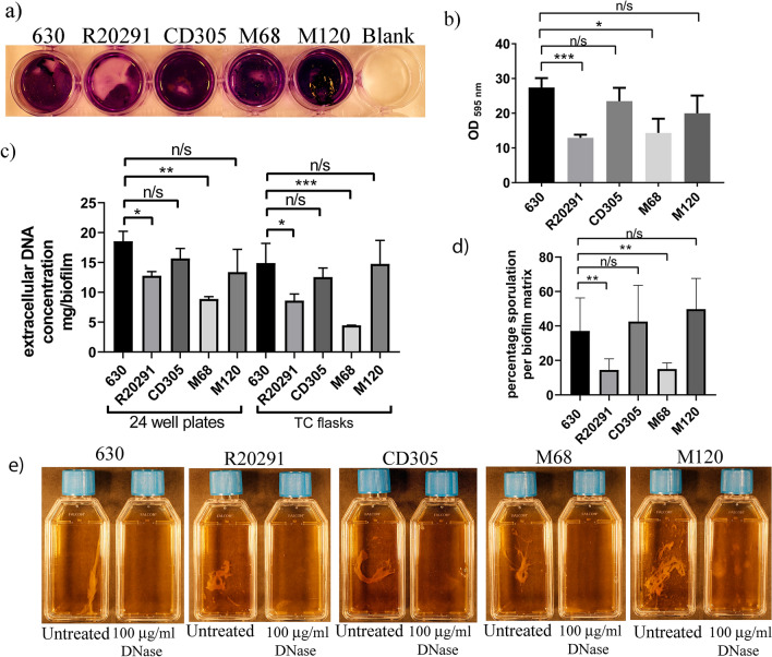
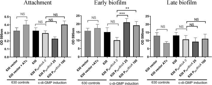
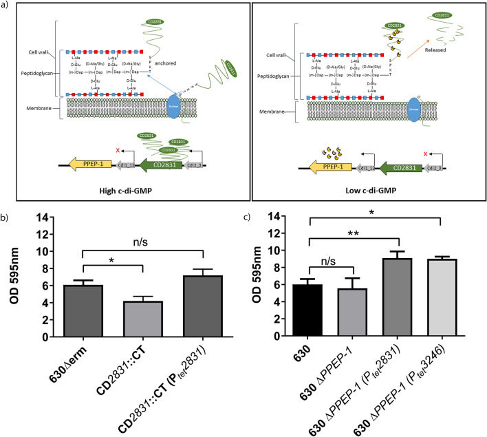
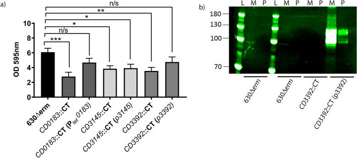
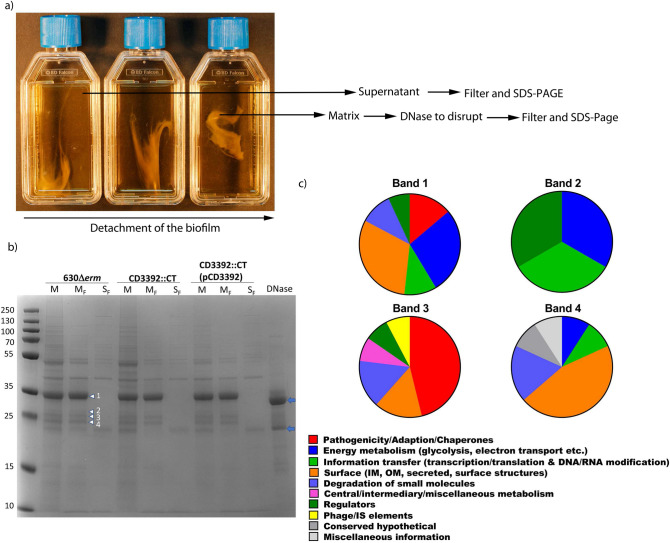
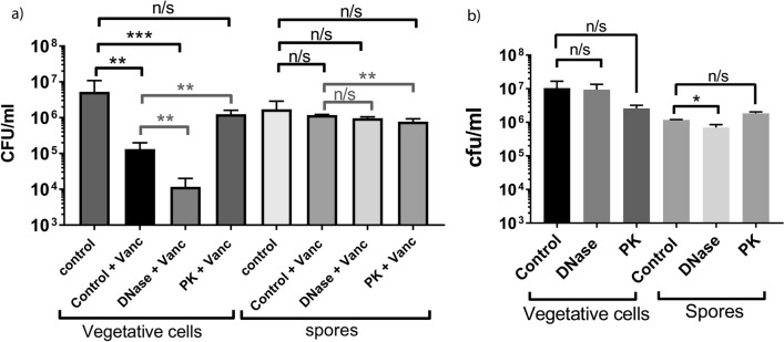
Clostridioides difficile is the leading cause of nosocomial antibiotic-associated diarrhoea worldwide, yet there is little insight into intestinal tract colonisation and relapse. In many bacterial species, the secondary messenger cyclic-di-GMP mediates switching between planktonic phase, sessile growth and biofilm formation. We demonstrate that c-di-GMP promotes early biofilm formation in C. difficile and that four cell surface proteins contribute to biofilm formation, including two c-di-GMP regulated; CD2831 and CD3246, and two c-di-GMP-independent; CD3392 and CD0183. We demonstrate that C. difficile biofilms are composed of extracellular DNA (eDNA), cell surface and intracellular proteins, which form a protective matrix around C. difficile vegetative cells and spores, as shown by a protective effect against the antibiotic vancomycin. We demonstrate a positive correlation between biofilm biomass, sporulation frequency and eDNA abundance in all five C. difficile lineages. Strains 630 (RT012), CD305 (RT023) and M120 (RT078) contain significantly more eDNA in their biofilm matrix than strains R20291 (RT027) and M68 (RT017). DNase has a profound effect on biofilm integrity, resulting in complete disassembly of the biofilm matrix, inhibition of biofilm formation and reduced spore germination. The addition of exogenous DNase could be exploited in treatment of C. difficile infection and relapse, to improve antibiotic efficacy.


Conflict of interest statement

The authors declare that the research was conducted in the absence of any commercial or financial relationships that could be construed as a potential conflict of interest.

Figures

Figure 1
Figure 1
A schematic and SEM visualisation of biofilm formation of C. difficile strain 630. (a) visualization of three stages of biofilm formation grown on coverslips in 24-well plates for 16 h (attachment), 24 h (early biofilm) and 72 h (late biofilms) prior to visualization. Surface attached appendages are indicated with an arrow, within the white box. White boxes indicate high magnification images present in supplementary data (Supplementary Fig. S1 online). Scale bar 5 µm. (b,c) Comparison of late biofilms formed on coverslips in 24 well plates and in tissue culture flasks, fixed and transferred to a stub for imaging. (b) Scale bar 10 µm (×1000), 5 µm (×3500 and ×5000), (c) scale bar 100 µm (×150), 10 µm (×1000 and ×3000).
Figure 2
Figure 2
Biofilm formation and eDNA concentrations of representative strains from each of the five C. difficile lineages. (a) Biofilm biomass determined by crystal violet assays performed in 24-well plates. (b) Quantification of biofilm biomass by methanol extraction of crystal violet assays performed in 24-well plates. Crystal violet assays are representative of six independent biological replicates. (c) Concentration of eDNA (C. difficile cells and spores were removed prior to eDNA quantification by filtering the vortexed disrupted biofilm samples) from duplicate biofilms produced in 24-well plates compared to TC flasks in a representative strain of all five C. difficile lineages: 630 (RT012), R20291 (RT027), CD305 (RT023), M68 (RT017) and M120 (RT078). (d) Percentage sporulation in the biofilm matrix produced in TC flasks for each strain in biological triplicates. (e) Duplicate late biofilm cultures were grown in tissue culture flasks, then detached by gentle agitation from the bottom of the tissue culture flask. One was left untreated and the other treated with 100 µg/mL DNase for 15 min to disrupt the biofilm matrix. Images of the tissue culture flasks were taken with a Cannon 600D SLR (mounted on a copy stand with lighting unit (Kaiser RS2) with a 50 mm prime lens). This was undertaken for C. difficile strains from all five lineages: 630 (RT012), R20291 (RT027), M120 (RT078), M68 (RT017) and CD305 (RT023). All error bars are SD. Statistical differences were assessed using linear regression analysis and significant differences are indicated *p < 0.05, **p < 0.01, ***p < 0.001.
Figure 3
Figure 3
The effect of the secondary messenger c-di-GMP on biofilm formation for C. difficile strain 630. In-vitro attachment, early biofilm and late biofilm formation in 24 well plates were quantified using crystal violet assays with strains of C. difficile 630 containing an empty plasmid (630 vector), or with a plasmid-based diguanylate cyclase (dccA) under control of a Ptet (inducible) promoter. These were compared to a 630 control strain containing a plasmid in the presence of 100 ng/mL anhydrotetracycline (630 vector + ATc). Biofilm and attachment assays were performed with a minimum of biological quadruplicate. The concentration of ATc used to induce dccA is listed in the graphs (0, 25 or 100 ng/mL), strain labelled 630Ptet dccA 0 is an uninduced vector control. Error bars are SD. Significant differences are calculated using linear regression analysis compared to the uninduced control strain 630Ptet dccA 0, **p < 0.01, ***p < 0.001.
Figure 4
Figure 4
The effect of c-di-GMP regulated cell wall proteins on biofilm formation for C. difficile strain 630. (a) Schematic showing the sortase-dependent anchoring of CD2831 to the peptidoglycan of C. difficile. Under high levels of c-di-GMP, CD2831 is expressed by the permissive interaction of c-di-GMP with the upstream type II riboswitch (Cdi2_3). The protease PPEP-1 is repressed under high levels of c-di-GMP due to the upstream type I riboswitch (Cdi1_12). Under low levels of c-di-GMP, expression of CD2831 is repressed and PPEP-1 is upregulated, resulting in the cleavage of CD2831 (and CD3246) from the peptidoglycan and release into the supernatant. This scheme is based on published data,,. (b) Crystal violet quantification of early biofilm formation produced in 24 well plates with an insertional inactivation mutant of CD2831 compared to its wildtype and inducible CD2831complement under control of anhydrotetracycline (ATc). (c) Crystal violet quantification of early biofilm formation produced in 24 well plates with overexpression of CD2831 and CD3246 under the control of an ATc inducible promoter, in an in-frame deletion mutant of the protease PPEP-1. Crystal violet assays were performed with a minimum of 6 biological replicates. Error bars are SD. Significant differences are calculated using linear regression analysis compared to all strains against its respective parent strain 630Δerm or 630, *p < 0.05, **p < 0.01.
Figure 5
Figure 5
Cell wall proteins involved in biofilm formation. (a) Insertional inactivation mutants of non-c-di-GMP regulated proteins CD0183, CD3145 and CD3392 were created in C. difficile strain 630Δerm. Mutants were complemented by introduction of plasmids expressing the wild type gene. These mutants and their complements were assessed for their ability to form early biofilms (24 h) on an abiotic surface in vitro compared to a wild-type control. Crystal violet assays were performed with a minimum of 6 biological replicates. All error bars are SD. Statistical differences were assessed using linear regression to compared to all strains against their respective parent strain 630Δerm, *p < 0.05, **p < 0.01, ***p < 0.001. (b) Western blot using anti CD3392 antibodies of the matrix (M) and planktonic fractions (P) of a late biofilm, from strains 630Δerm, CD3392::CT and CD3392 complement (CD3392::CT (pCD3392)). The biofilm matrix was detached from the bottom of a TC flask, disrupted by the addition of DNase (100 µg/mL), then loaded onto an SDS-PAGE gel alongside a protein ladder (L) for analysis by Western blot.
Figure 6
Figure 6
Analysis of the biofilm matrix and planktonic fractions. (a) Separation of the biofilm matrix and supernatant (planktonic fraction). After the biofilm matrix was detached from the bottom of the TC flask, the biofilm matrix was disrupted by the addition of DNase (100 µg/mL). (b) SDS-PAGE electrophoresis of the Biofilm matrix after DNase processing (M), filtered biofilm matrix (MF), compared to the filtered planktonic fraction (SF), and DNase control. The arrows on the left label the bands (1–4 top to bottom) sent for LC–MS/MS from strain 630∆erm, the blue arrows indicate the major and minor DNase band within the DNase control lanes. (c) Protein classification of the bands 1–4 presented in pie charts, with the percentage normalized total spectra. Colour codes indicate different functional classifications in accordance with the Riley classification.
Figure 7
Figure 7
The effect of vancomycin on CFU counts in disrupted and intact C. difficile strain 630 biofilms. (a) The effect of vancomycin treatment alone or in combination with DNase and Proteinase K on both vegetative cells and spores within an intact and degraded biofilm was undertaken in 3-day old biofilms, compared to untreated 3-day old biofilms. (b) A comparison between the effects of either DNase or proteinase K on the viability of vegetative cells and spores within a biofilm. The biofilms were detached from the bottom of the flask by gentle agitation and pre-treated with either vancomycin, or vancomycin supplemented with recombinant DNase or Proteinase K, compared to an untreated intact biofilm. Total counts and spores were enumerated and differentiated by heat inactivation of spore samples, thus killing the vegetative cells, enumerating spores alone. Experiments were undertaken with a minimum of 3 biological replicates. The data was analysed in Excel and GraphPad Prism 7.0, and error bars represent SD. Statistical analysis to determine the effect of vancomycin on disrupted and intact biofilm was performed using linear regression, **p < 0.01 (grey) and the effect of vancomycin and vancomycin combined with DNase and Proteinase K compared to untreated biofilms *p < 0.05, **p < 0.01, ***p < 0.001 (black).

Similar articles

Cited by

References

    1. Dawson LF, Valiente E, Wren BW. Clostridium difficile—a continually evolving and problematic pathogen. Infect. Genet. Evol. 2009;9:1410–1417. doi: 10.1016/j.meegid.2009.06.005. - DOI - PubMed
    1. Sorg JA, Sonenshein AL. Bile salts and glycine as cogerminants for Clostridium difficile spores. J. Bacteriol. 2008;190:2505–2512. doi: 10.1128/JB.01765-07. - DOI - PMC - PubMed
    1. Janoir C. Virulence factors of Clostridium difficile and their role during infection. Anaerobe. 2016;37:13–24. doi: 10.1016/j.anaerobe.2015.10.009. - DOI - PubMed
    1. Vardakas KZ, et al. Treatment failure and recurrence of Clostridium difficile infection following treatment with vancomycin or metronidazole: A systematic review of the evidence. Int. J. Antimicrob. Agents. 2012;40:1–8. doi: 10.1016/j.ijantimicag.2012.01.004. - DOI - PubMed
    1. Johnson S. Recurrent Clostridium difficile infection: A review of risk factors, treatments, and outcomes. J. Infect. 2009;58:403–410. doi: 10.1016/j.jinf.2009.03.010. - DOI - PubMed

Publication types

MeSH terms

LinkOut - more resources