. 2021 Feb 5;11(1):3263.
doi: 10.1038/s41598-021-82944-4.

Domeless receptor loss in fat body tissue reverts insulin resistance induced by a high-sugar diet in Drosophila melanogaster

Affiliations

Domeless receptor loss in fat body tissue reverts insulin resistance induced by a high-sugar diet in Drosophila melanogaster

Fernanda Lourido et al. Sci Rep. .

Abstract

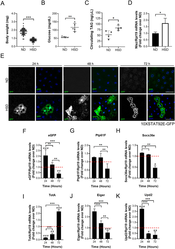
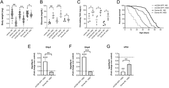
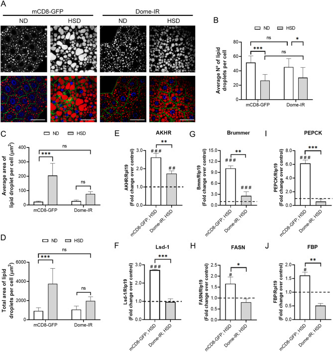
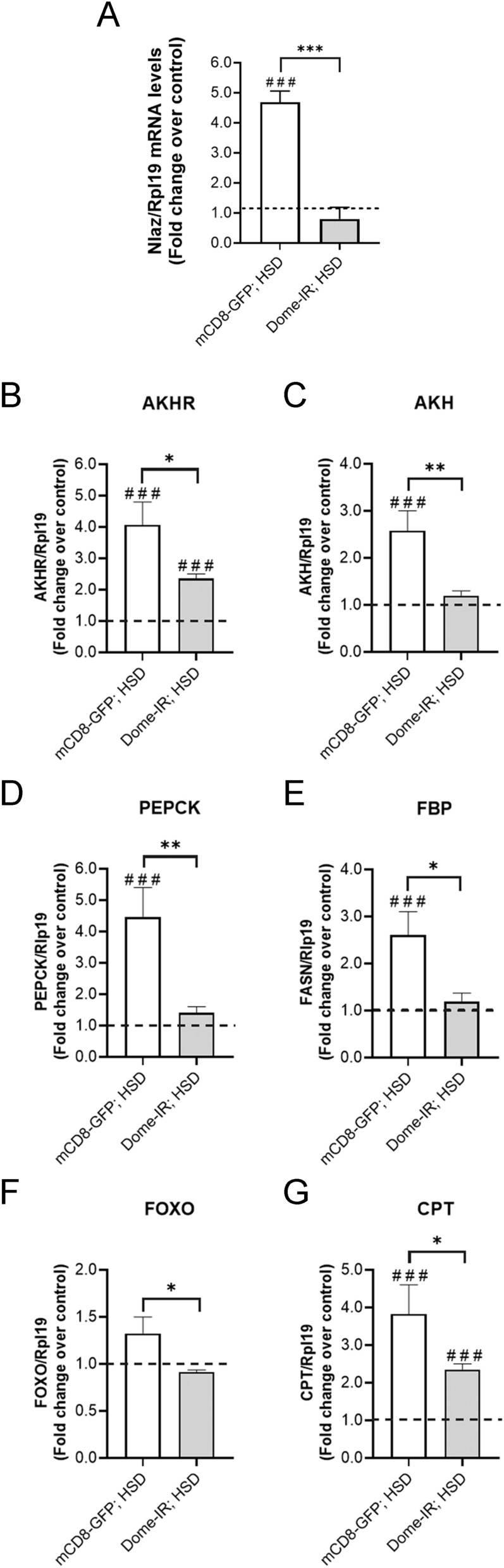
Insulin resistance is a hallmark of type 2 diabetes resulting from the confluence of several factors, including genetic susceptibility, inflammation, and diet. Under this pathophysiological condition, the dysfunction of the adipose tissue triggered by the excess caloric supply promotes the loss of sensitivity to insulin at the local and peripheral level, a process in which different signaling pathways are involved that are part of the metabolic response to the diet. Besides, the dysregulation of insulin signaling is strongly associated with inflammatory processes in which the JAK/STAT pathway plays a central role. To better understand the role of JAK/STAT signaling in the development of insulin resistance, we used a simple organism, Drosophila melanogaster, as a type 2 diabetes model generated by the consumption of a high-sugar diet. In this model, we studied the effects of inhibiting the expression of the JAK/STAT pathway receptor Domeless, in fat body, on adipose metabolism and glycemic control. Our results show that the Domeless receptor loss in fat body cells reverses both hyperglycemia and the increase in the expression of the insulin resistance marker Nlaz, observed in larvae fed a high sugar diet. This effect is consistent with a significant reduction in Dilp2 mRNA expression and an increase in body weight compared to wild-type flies fed high sugar diets. Additionally, the loss of Domeless reduced the accumulation of triglycerides in the fat body cells of larvae fed HSD and also significantly increased the lifespan of adult flies. Taken together, our results show that the loss of Domeless in the fat body reverses at least in part the dysmetabolism induced by a high sugar diet in a Drosophila type 2 diabetes model.


Conflict of interest statement

The authors declare no competing interests.

Figures

Figure 1
Figure 1
A high-sugar diet promotes JAK/STAT signaling activation in fat body cells. 10XSTAT92E-GFP larvae were reared on ND and HSDs. 72 h ND and 120 h HSD fed larvae (A) Bodyweight (n ≥ 20 animals at least 3 independent experiments), (B) Hemolymph glycemia (n = 70 larvae per pool, N ≥ 3 independent experiments), (C) Hemolymph TAG levels (n = 70 larvae per pool, N ≥ 3 independent experiments) and (D) Relative expression by qPCR to Nlaz in fat body cells (n = 25 larvae per pool, N ≥ 3 independent experiments). Bars represent mean ± SD. An unpaired two-tailed t-test was used to derive p value. ***p < 0.001, **p < 0.01, *p < 0.05 (E) Representative images of 10XSTAT92E-GFP reporter activations in fat body cells at 24, 48 and 72 h in 10XSTAT92E-GFP fat body larvae fed with normal (ND) or high-sugar diet (HSD). The panel shows cells marked with DAPI (blue), which expressed eGFP (green) when JAK/STAT signaling was activated. Scale bar = 50 µm. Relative expression by qPCR to eGFP (F), ptp61F (G), socs36E (H), totA (I), eiger (J), and upd2 (K) in fat body cells at 24, 48, and 72 h in larvae fed with an ND or HSD. The dotted line (red) represents the relative expression on larvae fed with ND (n = 25 animals per group, N = 3 independent experiments). 2-way ANOVA followed by Bonferroni's multiple comparisons test was used to derive all p values. ***p < 0.001, **p < 0.01, *p < 0.05. ###p < 0.001, ##p < 0.01, #p < 0.05 respect to ND control.
Figure 2
Figure 2
Specific dome knockdown in fat body cells reverses the effects of a high-sugar diet. Canton-S, control (mCD8-GFP), and Dome knockdown (Dome-IR) larvae were reared on ND for 72 h and HSD for 120 h. (A) Bodyweight (n ≥ 30 animals at least 4 independent experiments), (B) Hemolymph glycemia (n = 70 larvae per pool, N ≥ 3 independent experiments) and (C) Hemolymph TAG levels (n = 70 larvae per pool, N ≥ 3 independent experiments) of larvae fed with ND or HSD. (D) Lifespan curve of Dome-IR and mCD8-GFP adult male flies fed a ND or HSD (n = 300 animals per conditions). Relative expression by qPCR to dilp2 in brain cells (E), dilp6 (F), and dilp2 (G) in fat body cells of Dome-IR and mCD8-GFP larvae in HSD (n = 25 per group, N = 3 independent experiments). The dotted line (black) represents the relative expression by qPCR of mCD8-GFP and Dome-IR larvae fed with ND. Bars represent mean ± SD. 2-way ANOVA followed by Bonferroni's multiple comparisons test was used to derive all p values. ***p < 0.001, **p < 0.01, *p < 0.05. ###p < 0.001, ##p < 0.01, #p < 0.05 respect to ND control.
Figure 3
Figure 3
Dome loss in fat body cells reverses HSD effects on synthesis or lipid store. Control (mCD8-GFP) and Dome knockdown (Dome-IR) larvae were reared on ND for 72 h and HSD for 120 h. (A) Representative image of lipid droplets in fat body cells in different experimental conditions. The panel shows plasmatic membrane by mCD8-GFP expression (green), Nile Red stain (red), and DAPI (blue). Scale bar = 50 µm. (B) Quantification of the number of lipid droplets per cell, (C) average area of lipid droplets, and (D) total area of lipid droplets per cell in Dome-IR and mCD8-GFP early third instar larvae in normal or high-sugar diets. (n = 12 animals per group, N = 3 independent experiments). Relative expression by qPCR to akhr (E), lsd-1 (F), Bmm (G), fasn (H), pepck (I), and fbp (J) in fat body cells of Dome-IR and mCD8-GFP larvae fed in HSD (n = 25 animals per group, N = 3 independent experiments). The dotted line (black) represents the relative expression by qPCR of mCD8-GFP and Dome-IR larvae fed with ND. Bars represent mean ± SD. 2-way ANOVA followed by Bonferroni's multiple comparisons test was used to derive all p values. ***p < 0.001, **p < 0.01, *p < 0.05. ###p < 0.001, ##p < 0.01, #p < 0.05 respect to ND control.
Figure 4
Figure 4
Dome loss in fat body reversed high-sugar diet effect on peripheral insulin resistance related-markers. Control (mCD8-GFP) and Dome knockdown (Dome-IR) larvae were reared on ND for 72 h and HSD for 120 h. Relative mRNA levels evaluated by qPCR to Nlaz (A) in fat body cells, lipid mobilization related-proteins AKHR (B) and AKH (C), gluconeogenesis related-enzymes, PEPCK (D) and FBP (E) and FOXO signaling proteins, FOXO (F) and CPT (G) in whole larvae tissue of Dome-IR and mCD8-GFP early third instar larvae fed with normal diet or a high-sugar diet (n = 25 animals per group, N = 3 independent experiments). The dotted line (black) represents the relative expression by qPCR of mCD8-GFP and Dome-IR larvae fed with ND. Bars represent mean ± SD. 2-way ANOVA followed by Bonferroni's multiple comparisons test was used to derive all p values. ***p < 0.001, **p < 0.01, *p < 0.05. ###p < 0.001, ##p < 0.01, #p < 0.05 respect to ND control.

Similar articles

Cited by

References

    1. World Health Organization . Global Report on Diabetes. Geneva: WHO; 2016.
    1. Shoelson SE, Herrero L, Naaz A. Obesity, inflammation, and insulin resistance. Gastroenterology. 2007;132:2169–2180. doi: 10.1053/j.gastro.2007.03.059. - DOI - PubMed
    1. Andrali SS, Smapley ML, Vanderford NL, Özcan S. Glucose regulation of insulin gene expression in pancreatic β-cells. Biochem. J. 2008;415:1–10. doi: 10.1042/BJ20081029. - DOI - PubMed
    1. Weiss, M., Steiner, D. F., & Philipson, L. H. Insulin Biosynthesis, Secretion, Structure, and Structure-Activity Relationships (2000). - PubMed
    1. MacDonald PE, Joseph JW, Rorsman P. Glucose-sensing mechanisms in pancreatic β-cells. Philos. Trans. R. Soc. B Biol. Sci. 2005;360:2211–2225. doi: 10.1098/rstb.2005.1762. - DOI - PMC - PubMed

Publication types

MeSH terms

LinkOut - more resources