. 2021 Feb 5;4(1):168.
doi: 10.1038/s42003-021-01695-0.

Second-generation lung-on-a-chip with an array of stretchable alveoli made with a biological membrane

Affiliations

Second-generation lung-on-a-chip with an array of stretchable alveoli made with a biological membrane

Pauline Zamprogno et al. Commun Biol. .

Abstract

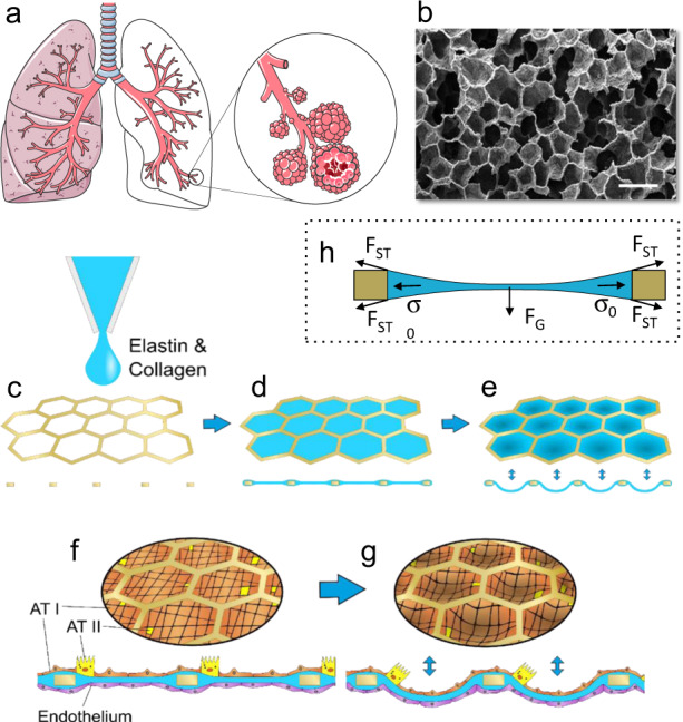
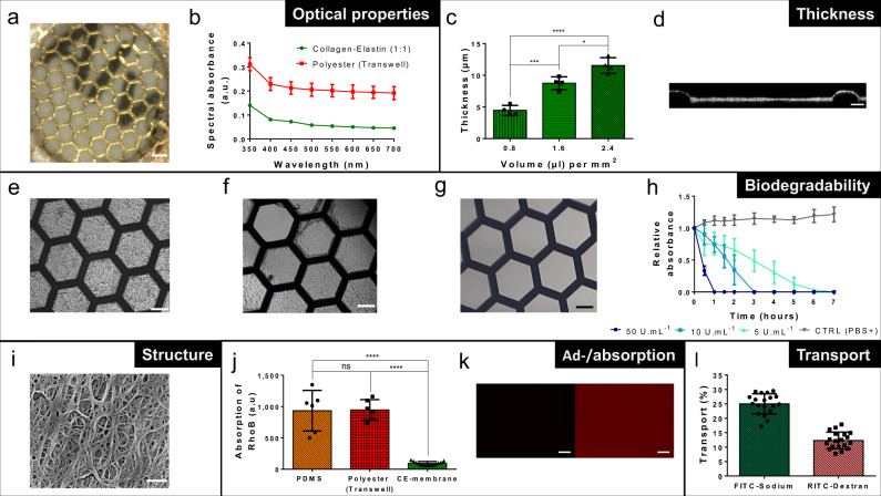
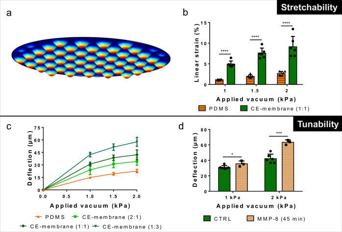
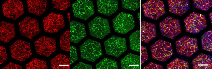
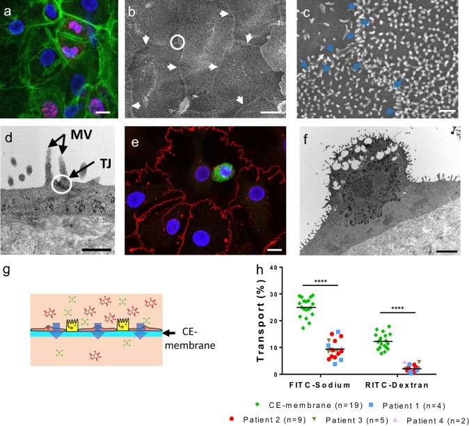
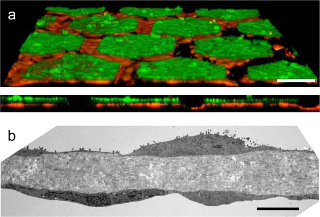
The air-blood barrier with its complex architecture and dynamic environment is difficult to mimic in vitro. Lung-on-a-chips enable mimicking the breathing movements using a thin, stretchable PDMS membrane. However, they fail to reproduce the characteristic alveoli network as well as the biochemical and physical properties of the alveolar basal membrane. Here, we present a lung-on-a-chip, based on a biological, stretchable and biodegradable membrane made of collagen and elastin, that emulates an array of tiny alveoli with in vivo-like dimensions. This membrane outperforms PDMS in many ways: it does not absorb rhodamine-B, is biodegradable, is created by a simple method, and can easily be tuned to modify its thickness, composition and stiffness. The air-blood barrier is reconstituted using primary lung alveolar epithelial cells from patients and primary lung endothelial cells. Typical alveolar epithelial cell markers are expressed, while the barrier properties are preserved for up to 3 weeks.


Conflict of interest statement

O.T.G. and J.D.S. are co-authors of a patent that describes the use of the mesh as in vitro barrier and whose rights are with the University of Bern and AlveoliX AG. All other authors declare no competing interests.

Figures

Fig. 1
Fig. 1. Second-generation lung-on-a-chip: creation of the lung alveoli array.
a Schematic of the respiratory tree-like structure ending with alveolar sacs (adapted from https://smart.servier.com/smart_image/lungs-7/, https://smart.servier.com/smart_image/lungs-11/ and https://smart.servier.com/smart_image/lungs/; Servier Medical Art by Servier; https://creativecommons.org/licenses/by/3.0/). b SEM picture of a slice of human lung parenchyma with tiny lung alveoli and their ultrathin air–blood barrier (courtesy of Prof. Dr. Peter Gehr, Institute of Anatomy, University of Bern; scale bar: 500 µm). c, d Schematic of the production of the CE membrane used in the second generation lung-on-a-chip. A thin gold mesh with an array of hexagonal pores of about 260 µm is used as a scaffold, on which a drop of collagen–elastin solution is pipetted. eg The collagen–elastin gel forms a suspended thin membrane that can be stretched at the alveolar level by applying a negative pressure on the basolateral side of the membrane. f, g Type I (ATI) and type II (ATII) primary human lung alveolar epithelial cells are cocultured with lung endothelial cells on the thin collagen–elastin membrane. h Schematic of the force balance during the drying of the membrane. FST, FG and σo stand for surface tension force, gravity and residual stress, respectively.
Fig. 2
Fig. 2. Properties of the thin biological membrane.
a Optical clarity of a 10-µm-thin CE membrane integrated in the gold mesh. Scale bar: 200 µm. b Comparison of the spectral absorbance of the CE membrane (n = 4) and of a polyester membrane (Transwell insert 0.4 µm pores sizes) (n = 3). c Characterization of the CE-membrane thickness in function of the collagen–elastin solution volume pipetted on top of the gold mesh (n = 4). d Cross-section of the CE membrane visualized via confocal microscopy. Scale bar: 20 µm. e Picture of an array of several hexagons with a CE membrane. Scale bar: 100 µm. f Local disruption (top left hexagon) of a membrane after being exposed to 10 U mL−1 MMP-8 in PBS+ during 1 h and stretched at −2 kPa. Scale bar: 100 µm. g Totally disrupted membrane after being exposed during 1 h to 50 U mL−1 of MMP-8 in PBS+. Scale bar: 100 µm. h CE-membrane degradation in function of the time and of the MMP-8 concentrations at 550 nm (n = 4). i SEM picture of the collagen and elastin fibers of the CE membrane. Scale bar: 500 nm. j Difference of rhodamine B (10 µM) absorption between a 10-µm-thin CE membrane (n = 13), a 10-µm-thin PDMS membrane (n = 6) and a polyester porous membrane (Transwell insert, 0.4 µm pores sizes) (n = 6). k Pictures of CE membrane (left) and a PDMS membrane (right) after being exposed to RhoB for 2 h. Scale bar: 200 µm. l Transport of FITC–sodium and RITC–dextran molecules across the CE membrane (n = 19).
Fig. 3
Fig. 3. Membrane flexibility.
a Numerical simulation of the deflection of the CE-membrane array. b Linear strain inside the CE membrane in function of the applied vacuum (n = 6). A 10-µm-thin PDMS membrane was taken as reference (n = 6). c Deflection of CE membranes of various compositions in function of an applied vacuum (n = 6 for CE membrane (1:1) and CE membrane (2:1) and n = 4 for CE membrane (1:3)). A 10-µm-thin PDMS membrane was taken as reference (n = 6). d Deflection of CE membrane exposed to MMP-8 in function of an applied vacuum (n = 6 for CE membrane (1:1) (CTRL) and n = 3 for treated membranes (MMP-8 (45 min))).
Fig. 4
Fig. 4. Immunostaining of primary human lung alveolar epithelial cells.
hAEpC cultured on the hexagonal mesh with the CE membrane after 4 days and at air–liquid interface for 2 days with expression of adherent junction markers (E-Cadherin, red), tight junctions with zonula occludens-1 (ZO-1, green) and merged (Hoechst, blue; E-Cadherin, red; ZO-1, green). Scale bar: 100 µm.
Fig. 5
Fig. 5. Primary lung alveolar epithelial cells.
a Expression of Ki-67 marker on hAEpC at day 4. Actin (green), Ki-67 (red) and Hoechst (blue). Scale bar: 10 µm. b SEM picture of hAEpC at day 14, illustrating tight cell–cell contacts. White arrows: cells border; white circle: area zoomed in c. Scale bar: 10 µm. c Intersection between three cells at day 14, showing their interface and a multitude of microvilli. Blue arrows: cells border. Scale bar: 1 µm. d TEM picture of tight junction (TJ) between two hAEpC. Apical microvillis (MV) typical to type II alveolar epithelial cells can clearly be seen. Scale bar: 500 nm. e Expression of surfactant protein-C (SP-C, green), tight junction (Z0-1, red) and nuclei (Hoechst, blue) at day 4. Scale bar: 10 µm. f TEM picture of a hAEpC type II-like cell at day 4, showing its microvilli and empty spaces, where lamellar bodies were located. Scale bar: 2 µm. g Schematic of the transport of molecules across the CE membrane cultured with alveolar epithelial cells. h Transport of FITC–sodium and RITC–dextran molecules across the CE membrane (n = 19) after 4 h of incubation with hAEpC the experiments were carried with cells from four patients (n = 20).
Fig. 6
Fig. 6. Air–blood barrier reproduction.
a Confocal pictures (perspective view and cross-section) of a coculture of hAEpC (E-Cadherin in green) with human primary endothelial cells (Rfp-label in red) on the hexagonal mesh with the CE membrane. Scale bar: 100 µm. b TEM picture of hAEpC type I-like cells in coculture with human lung endothelial cells at day 4. Scale bar: 5 µm.

References

    1. Huh D, Torisawa Y, Hamilton GA, Kim HJ, Ingber DE. Microengineered physiological biomimicry: organs-on-chips. Lab Chip. 2012;12:2156. doi: 10.1039/c2lc40089h. - DOI - PubMed
    1. Bhatia SN, Ingber DE. Microfluidic organs-on-chips. Nat. Biotechnol. 2014;32:760–772. doi: 10.1038/nbt.2989. - DOI - PubMed
    1. Ahadian S, et al. Organ-on-a-chip platforms: a convergence of advanced materials, cells, and microscale technologies. Adv. Healthc. Mater. 2018;7:14. doi: 10.1002/adhm.201700506. - DOI - PubMed
    1. Chan CY, et al. Accelerating drug discovery via organs-on-chips. Lab Chip. 2013;13:4697–4710. doi: 10.1039/c3lc90115g. - DOI - PMC - PubMed
    1. Esch EW, Bahinski A, Huh D. Organs-on-chips at the frontiers of drug discovery. Nat. Rev. Drug Discov. 2015;14:248–260. doi: 10.1038/nrd4539. - DOI - PMC - PubMed

Publication types

MeSH terms

Substances