Review
. 2021 Apr;41(4):707-717.
doi: 10.1038/s41372-021-00929-7. Epub 2021 Feb 5.

Modern pulmonary imaging of bronchopulmonary dysplasia

Affiliations
Review

Modern pulmonary imaging of bronchopulmonary dysplasia

Nara S Higano et al. J Perinatol. 2021 Apr.

Abstract

Bronchopulmonary dysplasia (BPD) is a complex and serious cardiopulmonary morbidity in infants who are born preterm. Despite advances in clinical care, BPD remains a significant source of morbidity and mortality, due in large part to the increased survival of extremely preterm infants. There are few strong early prognostic indicators of BPD or its later outcomes, and evidence for the usage and timing of various interventions is minimal. As a result, clinical management is often imprecise. In this review, we highlight cutting-edge methods and findings from recent pulmonary imaging research that have high translational value. Further, we discuss the potential role that various radiological modalities may play in early risk stratification for development of BPD and in guiding treatment strategies of BPD when employed in varying severities and time-points throughout the neonatal disease course.


Conflict of interest statement

CONFLICTS OF INTEREST

None

Figures

Figure 1:
Figure 1:
Clinical imaging of a patient with severe BPD and born extremely preterm (24 weeks gestation), acquired at ~41 weeks corrected gestational age. Left: Clinical chest x-ray radiograph (XR; coronal view). Right: x-ray computed tomography (CT; three axial views) of an extremely-preterm patient with BPD (24 weeks gestation at birth). XR and CT imaging occurred 4 days apart.
Figure 2:
Figure 2:
Lung ultrasound in B-mode. Left: normal lung ultrasound in infant without respiratory symptoms; normal lung artifacts. Right: lung ultrasound in an infant with severe BPD who requires mechanical ventilation for respiratory support; abnormal pleural line, interstitial lung fluid, hyperechogenicity, disruption of normal lung artifacts.
Figure 3:
Figure 3:
Comparison of slice-matched chest CT (left) and UTE MRI (right) in a neonatal patient with severe BPD. Despite the long interval between the two exams (77 days apart) and resulting progression of disease presentation, the image resolution and pulmonary tissue density are notably similar. Adapted from Higano NS, et al. J Magn Reson Imaging. 2017 Oct;46(4):992–1000 (52).
Figure 4:
Figure 4:
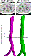
Top: Axial views of 3D respiratory-gated ultrashort echo-time (UTE) MRI in a neonate with tracheomalacia. Images were acquired during tidal breathing. Bottom: 3D surface renderings obtained from airway segmentation of 3D UTE images (left, pink, end-expiration; right, green, end-inspiration). From Bates AJ, et al. J Magn Reson Imaging. 2019 Mar;49(3):659–667 (71).
Figure 5:
Figure 5:
Hyperpolarized 3He gas ventilation MRI in a 2-month-old preterm infant. Coronal image views are shown. Various regions of ventilatory impairment are evident; the arrow denotes one such large defect. From Altes TA, et al. Clin Imaging. Sep-Oct 2017;45:105–110 (88).
Figure 6:
Figure 6:
Potential role of pulmonary imaging for preterm infants during their NICU course. The inpatient course is represented by two periods (perinatal through ~34 weeks corrected gestational age [CGA], and ~34 weeks CGA through discharge), with a more mild course and more severe course delineated on top and bottom, respectively. Status and recommended imaging modalities are represented by grey boxes, evidence based on clinical factors is represented by rounded, white boxes (solid border), and evidence based on tomographic imaging is represented rounded, white boxes (dotted border). The value and practicality of each modality will vary depending on time-point in disease progression, severity and co-morbidities in each patient, and capabilities of individual institutions. Abbreviations: CXR, chest x-ray radiograph; US, ultrasound; MRI, magnetic resonance imaging; CT, x-ray computed tomography; bronch, bronchoscopy; echo, echocardiography; cath, catheterization.

References

    1. Prayle A, Rosenow T. Looking under the bonnet of bronchopulmonary dysplasia with MRI. Thorax. 2020. February;75(2):100. - PubMed
    1. Raju TNK, Buist AS, Blaisdell CJ, Moxey-Mims M, Saigal S. Adults born preterm: a review of general health and system-specific outcomes. Acta Paediatr. 2017. September;106(9):1409–37. - PubMed
    1. Jobe AH. The new bronchopulmonary dysplasia. Curr Opin Pediatr. 2011. April;23(2):167–72. - PMC - PubMed
    1. Jobe AH, Bancalari E. Bronchopulmonary dysplasia. Vol. 163, American journal of respiratory and critical care medicine. United States; 2001. p. 1723–9. - PubMed
    1. Jensen EA, Wright CJ. Bronchopulmonary Dysplasia: The Ongoing Search for One Definition to Rule Them All. Vol. 197, The Journal of pediatrics. United States; 2018. p. 8–10. - PubMed

Publication types