Review
. 2021 Jun;17(6):369-385.
doi: 10.1038/s41581-021-00395-6. Epub 2021 Feb 5.

Planar cell polarity pathway in kidney development, function and disease

Affiliations
Review

Planar cell polarity pathway in kidney development, function and disease

Elena Torban et al. Nat Rev Nephrol. 2021 Jun.

Abstract

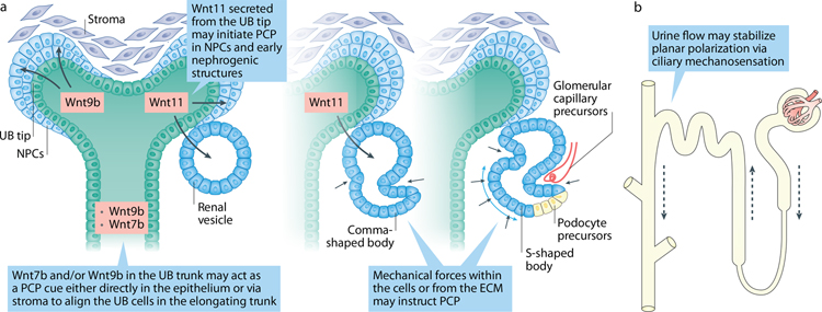
Planar cell polarity (PCP) refers to the coordinated orientation of cells in the tissue plane. Originally discovered and studied in Drosophila melanogaster, PCP is now widely recognized in vertebrates, where it is implicated in organogenesis. Specific sets of PCP genes have been identified. The proteins encoded by these genes become asymmetrically distributed to opposite sides of cells within a tissue plane and guide many processes that include changes in cell shape and polarity, collective cell movements or the uniform distribution of cell appendages. A unifying characteristic of these processes is that they often involve rearrangement of actomyosin. Mutations in PCP genes can cause malformations in organs of many animals, including humans. In the past decade, strong evidence has accumulated for a role of the PCP pathway in kidney development including outgrowth and branching morphogenesis of ureteric bud and podocyte development. Defective PCP signalling has been implicated in the pathogenesis of developmental kidney disorders of the congenital anomalies of the kidney and urinary tract spectrum. Understanding the origins, molecular constituents and cellular targets of PCP provides insights into the involvement of PCP molecules in normal kidney development and how dysfunction of PCP components may lead to kidney disease.


Figures

Figure 1:
Figure 1:. Planar cell polarity in Drosophila wing.
Left upper: Instructed by long-range cues, wing hexamer cells generate a single actin-based trichome (hair) at the most distal aspect of each cell. Right upper: Fat (Ft) mRNA is evenly expressed along the tissue, whereas Dachsous (Ds) transcript is expressed in a distal-proximal gradient and Four-jointed (Fj) kinase is expressed in a proximal-distal gradient. Fj phosphorylates both Ft (stronger phosphorylation at proximal side) and Ds (stronger phosphorylation at distal side). Strong Ft phosphorylation translates into a strong Ft-Ds protein-protein interactions at the proximal side, whereas Ds phosphorylation weakens Ft-Ds interactions at the distal part, thereby generating a shallow gradient of Ft-Ds activity across the tissue plane. Right middle: core PCP protein complexes are asymmetrically distributed: Vang-Pk to the proximal and Fz-Dsh-Dg to the distal sides. Cadherin Fmi is localized at both proximal and distal sides and forms homodimers between the extracellular domains of molecules expressed by adjacent cells. Interactions between Fmi and Vang or Fmi and Fz, as well as between extracellular domains of Vang and Fz expressed on surfaces of neighboring cells stabilize Vang-Pk and Fz-Dsh complexes on the opposite cell membranes. Inside the cell, mutual antagonism between Pk and Dsh creates “exclusion” zones where the proteins of the opposite core PCP complex cannot function. Right lower: Core PCP proteins control localization of PCP effectors via direct interactions (e.g. Vang interacts with In and Fy). In Drosophila wing cells, PCP effectors inhibit generation of trichome at the proximal side of the cell, whereas positive actin regulators, such as RhoA GTPase and Drosophila RhoA kinase (DROCK) accumulate at the distal side of the cell where they promote actin polymerization and hair formation.
Figure 2:
Figure 2:. Cell behaviors during morphogenesis
(A) Types of individual and collective cell behaviors that affect tissue architecture during morphogenesis. Cell elongation and oriented cell divisions act to promote tissue extension or orchestrate branching events. Cell intercalations are mediated by so called T1 transitions that involve four neighboring cells and by more complex intermediate ‘rosettes’ that include 5 or more cells. Apical constriction affects the curvature of the folding tissue and can promote neighbor cell exchanges. (B) Examples of cell behavior relevant to the formation of renal tubules. Both cell elongation and oriented cell divisions can stimulate tubule lengthening. Cell intercalations that accompany convergent extension rely on elongation and polarization of cells in the medio-lateral direction perpendicular to the tubular axis. Both cell intercalation and apical constriction reduce tubule diameter and lead to tubule elongation. Oriented cell divisions enable incorporation of a daughter cell along the tubular axis, facilitating tubule lengthening
Figure 3:
Figure 3:. PCP signaling in kidney development.
(A). Nephric/Wolffian duct formation is affected by mutations in certain PCP genes; the known mutated PCP genes are shown in red on the right side of each panel. Expression of PCP genes participating in the discussed process is depicted when known. (B). Outgrowth of ureteric bud from nephric duct is largely controlled by c-Ret (expressed in the ND at the time of UB formation) and its ligand GDNF (expressed in the cells of metanephric mesenchyme. Loss or mutations in several PCP genes lead to abnormal UB outgrowth in both human and mice resulting in renal agenesis or kidney duplication. (C). Ureteric bud branching morphogenesis depends on timely and spatially coordinated changes in cell shape and movements. Mutations in PCP genes affect UB branching, branch shape and branching angles. (D). Nephrogenic progenitor cell renewal and differentiation depend on the crosstalk between stroma and UB tip. Loss of stroma-expressing Fat4 or of NPC-expressing Dchs1/2 leads to NPC expansion. The specific signaling events involving Fat4/Dchs1–2 are unclear. (E). Podocyte foot processes are interdigitated in a precise fashion along the glomerular capillary. Loss of core PCP protein Vangl2 and Celrs1 affects podocyte differentiation, nephrin internalization and glomerular maturation.
Figure 4.
Figure 4.. Relationship between PCP and cystogenesis.
Various mechanisms contribute to cyst formation in renal tubules including loss of cilia and/or ciliary function, increased cell proliferation or abnormal apical-basal polarity. It was also proposed that lack of PCP protein function might contribute to cystogenesis. Accumulated experimental data have shown that core PCP proteins are polarized along the tubular axis in the developing kidneys, and that PCP signaling tightly controls the tubular diameter via CE and OCD. However, loss of core PCP proteins does not lead to cyst formation.
Figure 5.
Figure 5.. Potential instructive cues establishing PCP during kidney development.
The origin of PCP in early tubules is unknown, however, several molecules may potentially act as cues to initiate PCP in early nephrogenic structures. E.g. Wnt11 that expresses at the UB tip directs polarized NPC behaviors as they epithelize and transform into pre-tubular aggregates and renal vesicles. The link between apical-basal and PCP networks may coordinate establishment of planar polarization in the earliest structures. In the trunk, Wnt9b or Wnt7b may act locally in the UB trunk or non-autonomously on the cells adjacent to developing tubule to stabilize polarity of proliferating UB cells. Additionally, mechanical forces within the cells and/or from the ECM may provide polarity cues as comma- and S-shaped bodies undergo significant stretching and invagination. Onset of urine formation at E15.0 in the mouse nephron may provide further cues via ciliary mechanosensation to stabilize planar polarization along the growing tubule.

References

    1. Lawrence PA Gradients in the Insect Segment: The Orientation of Hairs in the Milkweed Bug Oncopeltus Fasciatus. Journal of Experimental Biology 44, 607–620 (1966). - PubMed
    1. Wong LL & Adler PN Tissue polarity genes of Drosophila regulate the subcellular location for prehair initiation in pupal wing cells. J Cell Biol 123, 209–221 (1993). - PMC - PubMed
    1. Gubb D & Garcia-Bellido A A genetic analysis of the determination of cuticular polarity during development in Drosophila melanogaster. J Embryol Exp Morphol 68, 37–57 (1982). - PubMed
    1. Adler PN The frizzled/stan pathway and planar cell polarity in the Drosophila wing. Curr Top Dev Biol 101, 1–31 (2012). - PMC - PubMed
    1. Vladar EK, Antic D & Axelrod JD Planar cell polarity signaling: the developing cell’s compass. Cold Spring Harb Perspect Biol 1, a002964 (2009). - PMC - PubMed

Publication types