. 2021 May;147(5):1355-1363.
doi: 10.1007/s00432-021-03542-5. Epub 2021 Feb 6.

Using ultrasound-targeted microbubble destruction to enhance radiotherapy of glioblastoma

Affiliations

Using ultrasound-targeted microbubble destruction to enhance radiotherapy of glioblastoma

Chanjuan Peng et al. J Cancer Res Clin Oncol. 2021 May.

Abstract

Objective: To investigate the efficacy and mechanism of ultrasound-targeted microbubble destruction (UTMD) combined with radiotherapy (XRT) on glioblastoma.

Methods: The enhanced radiosensitization by UTMD was assessed through colony formation and cell apoptosis in Human glioblastoma cells (U87MG). Subcutaneous transplantation tumors in 24 nude mice implanted with U87MG cells were randomly assigned to 4 different treatment groups (Control, UTMD, XRT, and UTMD + XRT) based on tumor sizes (100-300 mm3). Tumor growth was observed for 10 days after treatment, and then histopathology stains (HE, CD34, and γH2AX) were applied to the tumor samples. A TUNEL staining experiment was applied to detect the apoptosis rate of mice tumor samples. Meanwhile, tissue proteins were extracted from animal specimens, and the expressions of dsDNA break repair-related proteins from animal specimens were examined by the western blot.

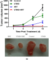
Results: When the radiotherapy dose was 4 Gy, the colony formation rate of U87MG cells in the UTMD + XRT group was 32 ± 8%, lower than the XRT group (54 ± 14%, p < 0.01). The early apoptotic rate of the UTMD + XRT group was 21.1 ± 3% at 48 h, higher than that of the XRT group (15.2 ± 4%). The tumor growth curve indicated that the tumor growth was inhibited in the UTMD + XRT group compared with other groups during 10 days of observation. In TUNEL experiment, the apoptotic cells of the UTMD + XRT group were higher than that of the XRT group (p < 0.05). The UTMD + XRT group had the lowest MVD value, but was not significantly different from other groups (p > 0.05). In addition, γH2AX increased due to the addition of UTMD to radiotherapy compared to XRT in immunohistochemistry (p < 0.05).

Conclusions: Our study clearly demonstrated the enhanced destructive effect of UTMD combined with 4 Gy radiotherapy on glioblastoma. This could be partly achieved by the increased ability of DNA damage of tumor cells.

Keywords: Glioblastoma; Microbubbles; Radiation therapy; Ultrasound; Ultrasound therapy.


Conflict of interest statement

The authors declare no conflict of interest.

Figures

Fig. 1
Fig. 1
Clonogenic survival and cell apoptosis in U87MG cells. a Clonogenic survival of different irradiation doses for the XRT and UTMD + XRT groups. Representative images showed the surviving colonies. b Graphs showed the changes of clonogenic survival fraction. **p < 0.01 versus XRT group. c Cell survival curve and click multi-target fitting curve. Data present average results from three independent experiments (n = 3). d Representative images showing apoptosis in U87MG cells at 48 h after treatment. Percentage of the bottom right quadrant showed the average value of early apoptosis
Fig. 2
Fig. 2
Effects of UTMD and radiation on the growth of U87MG xenografts in nude mice. a Tumor growth curves of different groups after treatment. b Photographs of excised xenografts from inoculated mice in different groups. Data are mean ± SD of the six mice per group (n = 6)
Fig. 3
Fig. 3
Histopathological findings of XRT combined with UTMD on tumor tissues. a Top row: the H&E staining of excited xenograft tissues showed typical glioblastoma with all otypic nuclear division and tumor giant cells; second row: representative images of CD34 stained tumour cross-sections, The scale bars represent 50 μm; Third row: representative images of apoptosis cells in tunnel experiment (× 100). b MVD (microvasculature density) comparison in different groups (n = 6). c Comparison of cell apoptosis in tissues of four groups of rats (n = 6), *p < 0.05 versus XRT groups
Fig. 4
Fig. 4
Western blot analysis showing the effects of UTMD combined with XRT on DSB repair-related proteins on xenograft tumor tissues. a Representative western blots showing the effects of UTMD on expression of ATM, ATR, CHK1, CHK2, H2AX, P53. b Graphs showing changes of protein expression in different groups. c Representative western blots showing the effects of UTMD on expression of Phospho-ATR, Phospho-BRCA1, Phospho-CHK1, Phospho-CHK2, γH2AX and Phospho-P53 (d) Graphs showing changes of protein expression among different groups. β-action was included as a loading control. Data represent average results from three independent experiments; SD signifies standard deviation (n = 3), *p < 0.05 versus XRT groups
Fig. 5
Fig. 5
γH2AX labeling of tumor sections. a Representative images of γH2AX stained tumour cross-sections, The scale bars represent 200 μm. b γH2AX labeling comparison in different groups (n = 6), *p < 0.05 versus XRT groups

References

    1. Alexander BM, Cloughesy TF (2017) Adult glioblastoma. J Clin Oncol 35(21):2402–2409 - PubMed
    1. Al-Mahrouki AA et al (2014) Cellular characterization of ultrasound-stimulated microbubble radiation enhancement in a prostate cancer xenograft model. Dis Model Mech 7(3):363–372 - PMC - PubMed
    1. Briggs K et al (2014) Non-invasive monitoring of ultrasound-stimulated microbubble radiation enhancement using photoacoustic imaging. Technol Cancer Res Treat 13(5):435–444 - PMC - PubMed
    1. Carruthers RD et al (2018) Replication stress drives constitutive activation of the DNA damage response and radioresistance in glioblastoma stem-like cells. Cancer Res 78(17):5060–5071 - PMC - PubMed
    1. Chabowski M et al (2018) Comparison of microvessel density using nestin and CD34 in colorectal cancer. Anticancer Res 38(7):3889–3895 - PubMed

LinkOut - more resources