. 2021 Mar;130(1):53-60.
doi: 10.1007/s00412-021-00751-2. Epub 2021 Feb 6.

Chromosome Y pericentric heterochromatin is a primary target of HSF1 in male cells

Affiliations

Chromosome Y pericentric heterochromatin is a primary target of HSF1 in male cells

Jessica Penin et al. Chromosoma. 2021 Mar.

Abstract

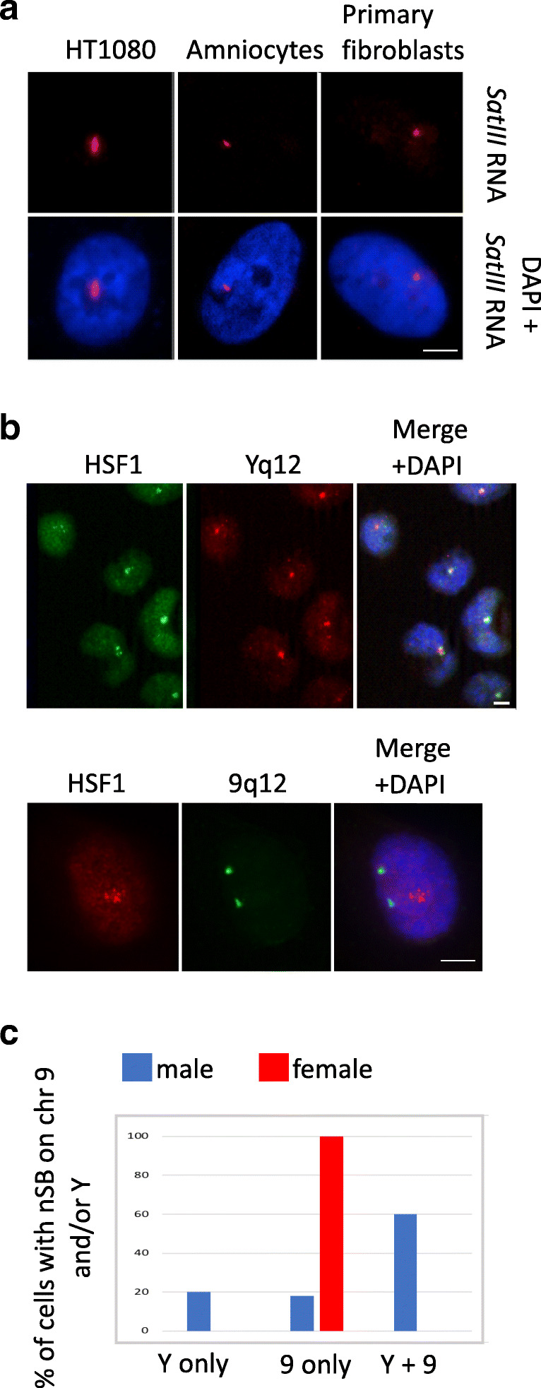
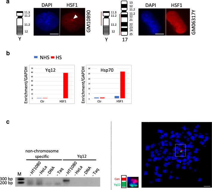
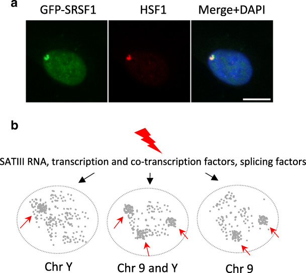
The heat shock factor 1 (HSF1)-dependent transcriptional activation of human pericentric heterochromatin in heat-shocked cells is the most striking example of transcriptional activation of heterochromatin. Until now, pericentric heterochromatin of chromosome 9 has been identified as the primary target of HSF1, in both normal and tumor heat-shocked cells. Transcriptional awakening of this large genomic region results in the nuclear accumulation of satellite III (SATIII) noncoding RNAs (ncRNAs) and the formation in cis of specific structures known as nuclear stress bodies (nSBs). Here, we show that, in four different male cell lines, including primary human fibroblasts and amniocytes, pericentric heterochromatin of chromosome Y can also serve as a unique primary site of HSF1-dependent heterochromatin transcriptional activation, production of SATIII ncRNA, and nucleation of nuclear stress bodies (nSBs) upon heat shock. Our observation suggests that the chromosomal origin of SATIII transcripts in cells submitted to heat shock is not a determinant factor as such, but that transcription of SATIII repetitive units or the SATIII ncRNA molecules is the critical element of HSF1-dependent transcription activation of constitutive heterochromatin.

Keywords: HSF1; Heterochromatin; Human; nSB; ncRNA.


Figures

Fig. 1
Fig. 1
Heat-shocked HT1080 cells display a unique HSF1 foci on chromosome Y. a Unique SATIII RNA foci are observed in the nucleus of cell lines of male origin (bar = 5 μm). SATIII RNA (red signals) are detected by RNA FISH with a fluorescent oligonucleotide recognizing (GGAAU)n motifs, on male cells of different origin (HT1080 cells, amniocytes and primary fibroblasts). b HSF1 detected by immuno-fluorescence in HT1080 cells colocalizes with the Yq12 locus and not with the 9q12 locus detected by DNA FISH. Upper image Yq12 locus (red signals), HSF1 (green signal) (bar = 5 μm). Lower image 9q12 locus (green signal), HSF1 (red signal) (bar = 5 μm). c Graph giving the percentage of nSBs present on chromosome 9, chromosome Y, or both, in normal fibroblasts. HSF1 foci were codetected by immunofluorescence and FISH in human primary stressed male and female fibroblasts (total of 200 nuclei analyzed on three independent analyses)
Fig. 2
Fig. 2
HSF1 overexpression leads to its binding to pericentric regions of chromosome 9 in HT1080 cells. a HT1080 cells do not lack chromosome-9-specific SATIII genomic sequences. Simultaneous detection of SATIII sequences from chromosome 9 pericentric regions detected with the pHuR98 probe (red signal) and of alphoid sequences from chromosome 9 centromeric regions with the pMR9A (green signal) on mitotic spreads from HT1080 cells. The two chromosomes 9 are also enlarged (bar = 5 μm). b HT1080 cells transiently transfected with a HSF1-GFP expression vector display HSF1 foci (green signals) at the 9q12 loci detected by FISH (red signal) (bar = 10 μm)
Fig. 3
Fig. 3
HSF1 binds chromosome Y SATIII sequences upon heat shock. a HSF1 detection by immunofluorescence on somatic hamster/human hybrid cell lines containing either chromosome Y (GM10890 cells) or only the Yp->Yq11::17q12->17qter (GM06317Y cells) region of chromosome Y. HSF1 foci were only detected in hybrid cell lines containing the Yq12 locus (bar = 10 μm). b ChIP experiments with anti-HSF1 antibody performed on unstressed (NHS) and heat shock (HS) HT1080 cells (anti-IgG antibody were used as a control, Ctr). PCR amplifications were performed with primers specific to either chromosome-Y-specific SATIII sequences or to the HSP70 gene. c The enrichment of genomic SATIII sequences of chromosome Y origin in anti-HSF1 ChIP experiments was assessed. First (left image), a PCR product, here analyzed by electrophoresis, was only obtained with genomic DNA from stressed HT1080 cell line of male origin and not with genomic DNA from stressed HeLa cells of female origin. Controls are shown in which the amplification reactions were performed with no DNA or no DNA polymerase (Taq). A PCR product is only obtained with genomic DNA of male origin. Second (right image), the PCR product obtained from HT1080 cells was labeled and used for a DNA FISH experiment on metaphase spreads from HT1080 cells (green signal). A centromeric probe was also used to identify chromosome Y (red signal) (bar = 5 μm). The PCR product hybridizes to chromosome Y when used as a probe in a DNA FISH experiment
Fig. 4
Fig. 4
SRSF1 is recruited to chromosome-Y-specific active SATIII transcription sites in human cells upon heat shock. a SRSF1-GFP splicing factors form unique foci (green signal) colocalizing with HSF1 (red signal) detected by immunofluorescence in stressed HT1080 cells (bar = 10 μm). b Model of formation or primary nSBs on either chromosome 9, chromosome Y, or both chromosomes 9 and Y. In stressed cell of male origin, pericentric regions of either chromosome 9, chromosome Y, or both are actively transcribed. The transcription of SATIII repetitive sequences at nSBs results in the titration of transcription and splicing factors from the rest of the nucleus

References

    1. Akerfelt M, Vihervaara A, Laiho A, et al. Heat shock transcription factor 1 localizes to sex chromatin during meiotic repression. J Biol Chem. 2010;285(45):34469–34476. doi: 10.1074/jbc.M110.157552. - DOI - PMC - PubMed
    1. Akerfelt M, Morimoto RI, Sistonen L. Heat shock factors: integrators of cell stress, development and lifespan. Nat Rev Mol Cell Biol. 2010;11:545–555. doi: 10.1038/nrm2938. - DOI - PMC - PubMed
    1. Antonacci R, Marzella R, Finelli P, Lonoce A, Forabosco A, Archidiacono N, Rocchi M. A panel of subchromosomal painting libraries representing over 300 regions of the human genome. Cytogenet Cell Genet. 1995;68:25–32. doi: 10.1159/000133882. - DOI - PubMed
    1. Barna J, Csermely P, Vellai T (2018) Roles of heat shock factor 1 beyond the heat shock response. Cell Mol Life Sci 75(16):2897–2916 - PMC - PubMed
    1. Biamonti G, Vourc’h C. Nuclear stress bodies. Cold Spring Harb Perspect Biol. 2010;2(6):a0000695. doi: 10.1101/cshperspect.a000695. - DOI - PMC - PubMed

Publication types

MeSH terms

LinkOut - more resources