Review
. 2021 Feb;15(1):21-35.
doi: 10.1007/s12072-020-10121-2. Epub 2021 Feb 6.

How does hepatic lipid accumulation lead to lipotoxicity in non-alcoholic fatty liver disease?

Affiliations
Review

How does hepatic lipid accumulation lead to lipotoxicity in non-alcoholic fatty liver disease?

Yana Geng et al. Hepatol Int. 2021 Feb.

Abstract

Background: Non-alcoholic fatty liver disease (NAFLD), characterized as excess lipid accumulation in the liver which is not due to alcohol use, has emerged as one of the major health problems around the world. The dysregulated lipid metabolism creates a lipotoxic environment which promotes the development of NAFLD, especially the progression from simple steatosis (NAFL) to non-alcoholic steatohepatitis (NASH).

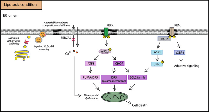
Purposeand aim: This review focuses on the mechanisms of lipid accumulation in the liver, with an emphasis on the metabolic fate of free fatty acids (FFAs) in NAFLD and presents an update on the relevant cellular processes/mechanisms that are involved in lipotoxicity. The changes in the levels of various lipid species that result from the imbalance between lipolysis/lipid uptake/lipogenesis and lipid oxidation/secretion can cause organellar dysfunction, e.g. ER stress, mitochondrial dysfunction, lysosomal dysfunction, JNK activation, secretion of extracellular vesicles (EVs) and aggravate (or be exacerbated by) hypoxia which ultimately lead to cell death. The aim of this review is to provide an overview of how abnormal lipid metabolism leads to lipotoxicity and the cellular mechanisms of lipotoxicity in the context of NAFLD.

Keywords: Cell death; ER stress; Free fatty acids; JNK; Lipid metabolism; Lipotoxicity; MAFLD; Mitochondrial dysfunction; NAFLD; NASH.


Conflict of interest statement

There is no potential conflict of interest.

Figures

Fig. 1
Fig. 1
Hepatic lipid metabolism in NAFLD. Dietary lipids are mainly absorbed in the intestine and incorporated into intestinal chylomicrons (intestinal CMs) and then transported to adipose tissue and the liver via the bloodstream. In adipose tissue, intestinal CMs are taken up and stored in lipid droplet. In NAFLD, the hydrolysis of TG is largely enhanced in adipocytes. TG is hydrolyzed into diacylglycerol (DAG), monoacylglycerol (MAG) and glycerol, releasing one fatty acid in each step. The increased fatty acid load is then delivered to the liver, scavenged by fatty acid translocase (FAT/CD36), fatty acid transport proteins (FATPs) and caveolins and incorporated into lipid droplets. In hepatocytes, de novo lipogenesis is increased which contributes to the lipid pool as well. On the other hand, fatty acids are oxidized in mitochondria (mitochondrial β-oxidation), peroxisomes (peroxisomal β-oxidation) and microsomes (microsomal ω-oxidation), accompanied by an elevated production of ROS. The secretion of TG-enriched very-low-density lipoprotein (VLDL) is another route of lipid disposal. It is increased in absolute terms, but its increase does not match the increased fat accumulation in NAFLD. Arrows (red) indicate the up- or down-expressions of specific proteins in NAFLD. CPT1/2 carnitine palmitoyltransferase ½, FFA free fatty acid, TCA cycle: the citric acid cycle, ETC electron transport chain, FABP fatty acid-binding protein, ACC acetyl-CoA carboxylase, FAS fatty acid synthase, DGAT diacylglycerol acyltransferase, MGAT monoacylglycerol acyltransferase
Fig. 2
Fig. 2
Mechanisms of lipotoxicity in NAFLD. The mechanisms of lipotoxicity involve several cellular processes, such as ER stress, mitochondrial dysfunction, lysosomal dysfunction, the release of extracellular vesicles (EVs) and hypoxia, ultimately leading to cell death. Additionally, the activation of JNK signaling pathway also plays an important role in lipoapoptosis and its activation is closely associated with ER stress/UPR and mitochondrial dysfunction
Fig. 3
Fig. 3
Lipotoxic UPR induces hepatic cell death. Under lipotoxic condition, several factors are implicated in the occurrence of ER stress, including disrupted ER-to-Golgi protein trafficking, altered ER membrane composition and stiffness, impaired VLDL-TG assembly and the change of Ca2+ gradient by the decreased level of SERCA2 activity. PERK is activated and leads to the phosphorylation of eIF2α, subsequently causes the activation of downstream ATF3- and CHOP-related apoptotic signaling pathways. IRE1α could also be activated by toxic lipids, thus stimulates TRAF2/ASK1/JNK signaling pathway. On the other hand, spliced XBP1 mediates adaptive UPR, which aims to alleviate ER stress. Together with apoptotic UPR, the massive efflux of Ca2+ from ER could result in mitochondrial dysfunction and the initiation of apoptotic cell death. VLDL very-low-density lipoprotein, SERCA2 sarco/endoplasmic reticulum Ca2+-ATPase 2, ATF3 activating transcription factor 3, ASK1 apoptosis signal-regulating kinase 1, CHOP C/EBP homologous protein, DR5 death receptor 5, DP5 death protein 5, eIF2α eukaryotic initiation factor 2α, TRAF2 TNF receptor-associated factor 2, IRE1α inositol-requiring kinase 1α, JNK c-Jun N-terminal kinase, PERK double-stranded RNA-dependent protein kinase-like ER kinase, PUMA p53 upregulated modulator of apoptosis, sXBP1 spliced X-Box Binding Protein 1
Fig. 4
Fig. 4
Modes of cell death in NASH. Apoptosis, necroptosis and pyroptosis have all been described in NASH. Apoptosis, which can be demonstrated by caspase 3/7 activation, is triggered through both intrinsic and extrinsic pathways in NASH. Lysosomal dysfunction and ER stress are involved in the initiation of intrinsic pathway. In addition, caspase 2 is increased in NASH, whereas caspase 9 level is reduced in ballooned hepatocytes. In the extrinsic pathway, death receptor 5, apoptosis antigen 1 (Fas) and tumor necrosis factor receptor 1 (TNF-R1) are increased in NASH leading to activation of caspase 8 and induction of apoptosis. Necroptosis is characterized as a mixed lineage kinase domain-like pseudokinase (MLKL) related pore formation in the plasma membrane. It is induced by Receptor-Interacting Protein 3 (RIP3) and RIP1. Caspase 8 inhibits RIP3 activation. Pyroptosis is characterized as gasdermin D (GSDMD) mediated pore formation in the plasma membrane. Its activation is accompanied by inflammasome formation, which involves the maturation of caspase 1, IL-1β and NLRP3. Arrows (red) indicate the up- or down-expressions of specific proteins in NAFLD. IL-1β interleukin-1β, NLRP3 NACHT, LRR and PYD domains-containing protein 3

References

    1. Eslam M, et al. A new definition for metabolic associated fatty liver disease: an international expert consensus statement. J Hepatol. 2020;73:201. - PubMed
    1. Byrne CD, Perseghin G. Non-alcoholic fatty liver disease: a risk factor for myocardial dysfunction? J Hepatol. 2018;68(4):640–642. doi: 10.1016/j.jhep.2017.12.002. - DOI - PubMed
    1. Friedman SL, et al. Mechanisms of NAFLD development and therapeutic strategies. Nat Med. 2018;24(7):908–922. doi: 10.1038/s41591-018-0104-9. - DOI - PMC - PubMed
    1. Cholankeril G, et al. Liver transplantation for nonalcoholic steatohepatitis in the US: temporal trends and outcomes. Dig Dis Sci. 2017;62(10):2915–2922. doi: 10.1007/s10620-017-4684-x. - DOI - PubMed
    1. Younossi ZM, et al. Epidemiology of nonalcoholic fatty liver disease and nonalcoholic steatohepatitis: implications for liver transplantation. Transplantation. 2019;103(1):22–27. doi: 10.1097/TP.0000000000002484. - DOI - PubMed