Review
. 2021 Mar 17;109(6):918-937.
doi: 10.1016/j.neuron.2021.01.013. Epub 2021 Feb 5.

Unraveling circuits of visual perception and cognition through the superior colliculus

Affiliations
Review

Unraveling circuits of visual perception and cognition through the superior colliculus

Michele A Basso et al. Neuron. .

Abstract

The superior colliculus is a conserved sensorimotor structure that integrates visual and other sensory information to drive reflexive behaviors. Although the evidence for this is strong and compelling, a number of experiments reveal a role for the superior colliculus in behaviors usually associated with the cerebral cortex, such as attention and decision-making. Indeed, in addition to collicular outputs targeting brainstem regions controlling movements, the superior colliculus also has ascending projections linking it to forebrain structures including the basal ganglia and amygdala, highlighting the fact that the superior colliculus, with its vast inputs and outputs, can influence processing throughout the neuraxis. Today, modern molecular and genetic methods combined with sophisticated behavioral assessments have the potential to make significant breakthroughs in our understanding of the evolution and conservation of neuronal cell types and circuits in the superior colliculus that give rise to simple and complex behaviors.

Keywords: action; adaptation; attention; avoidance; conservation; decision-making; defensive behaviors; escape behaviors; evolution; lamination; motor maps; neural cartography; optic tectum; orienting; prey capture; sensory maps; vision.


Figures

Figure 1.
Figure 1.
The relative size of the SC varies across evolution. Cladogram of tree terminations showing schematic images of the brains of different species. The blue coloring highlights the SC indicating the relative size differences. Note that in mammals, the SC lies under the cerebral cortex. Brain images are not to scale.
Figure 2.
Figure 2.
The SC shows varying lamination and retinal axon origin. Nissl stained coronal sections through the left SC of six species showing differences in layer differentiation and laminar nomenclature. a. SC of monkey (Macaca mulatta); b. California grey squirrel (Sciurus griseus); c. OT of English sparrow (Passer domesticus); d. SC of mouse (Mus musculus). e. OT of the pigeon (Columba livia domestica) f. SC of the tree shew (Tupaia belangeri). Note that the layering is more pronounced in birds than mammals. a-d scale bars = 1mm. a-e from the PhD thesis of Daniel Major, images kindly provided by Dr. Harvey Karten. See text for abbreviations. g. Schematic diagram of the patterns of retinal afferents entering the OT superficially and terminating in different layers forming separate visual channels. h. Same as in g for the mammalian SC with retinal afferents entering the SC from below. Adapted with permission from (Yamagata et al., 2006).
Figure 3.
Figure 3.
The SC forms topographical maps of space and action. a. Schematic illustration of the right SC map from mouse (Mus musculus) and b. monkey (Macaca mulatta). In the monkey, the central 15° takes up much of the SC visual map whereas in mice, the nasal and temporal visual fields are represented uniformly across the SC. Adapted from (Robinson, 1972; Dräger and Hubel, 1976 and Cang et al., 2018 with permission).
Figure 4.
Figure 4.
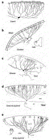
Wide field vertical (WFV) neuron morphology is conserved across species. a. Lizard (Anolis carolinensis), from (Ramón y Cajal, 1995). b. Chicken (Galllus gallus), from (Luksch et al., 1998). c. Mouse (Mus musculus), from (Masterson et al., 2019). d. Ground squirrel (Spermophilius beecheyi), from (Major et al., 2000). e. Grey squirrel (Sciurus carolinensis) from (May, 2006). Images adapted with permission.
Figure 5.
Figure 5.
The mammalian SC and avian OT influence the entire neuraxis through their extensive inputs and outputs. A selected sample of inputs appear on the left and outputs on the right. Orange shows intermediate and deeper layer inputs and outputs and blue shows superficial layer inputs and outputs. Tectopontine refers to uncrossed pathways. Tectobulbar refers to crossed pathways. Adapted with permission from (Luksch, 2003).
Figure 6.
Figure 6.
Schematic of retino-isthmo-tectal circuit. Shepard’s crook neurons (red) in the OT project to the Ipc nucleus and the Ipc neurons (blue) in turn, provide cholinergic (ACh) feedback to the OT. Retinal terminals (black arising from the top layer 1) make synaptic contacts with dendrites of OT tectal ganglion neurons (grey discs and complex dendritic fields). The GABAergic input from the Imc is widespread (green). Note that the circuit depicted in red, blue and green is repeated throughout the OT but only one section is highlighted here for clarity. Adapted with permission from (González-Cabrera et al., 2016).
Figure 7.
Figure 7.
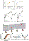
SC neuronal activity encodes saccade choice. a. Schematic of the odd-ball target selection task in which a monkey correctly chose the red target on all trials. Black lines show saccade trajectories. b. After muscimol injection into the SC, monkeys made more frequent errors selecting the distractor stimuli. c. During recovery, 24 hours after muscimol, monkeys performed accurately again. Adapted from (McPeek and Keller, 2004). d. Schematic of the odd-ball target selection task in which a monkey showed good (>75% accuracy) and e. poor (<75% accuracy) target selection performance. Black lines show saccade trajectories. Scale bars indicate 5°. f. Population responses to targets compared to distractors simulated from multiple SC neuron recordings, showed higher discriminability during good performance compared to g. poor performance. Warmer colors indicate higher spike rates. Adapted from (Kim and Basso, 2008).
Figure 8.
Figure 8.
The SC of monkeys plays a causal role in perceptual decision-making. a. Example proportion of ‘Yes’ responses of a monkey in the conservative priming trials (orange more ‘No” trials). Black shows equal “yes’ ‘No” trails (Baseline). b. Same as in a for trials with (orange) and without (black) stimulation of the SC, which mimicked behavioral changes in decision criterion stemming from changes in stimulus frequency. c - e. Neuronal activity from SC neurons during the decision epoch for “Yes’ (solid) and “No” (dashed) trials. After conservative priming the difference in neuronal activity decreased whereas after liberal priming the difference increased. Grey and black lines show different coherences. Adapted with permission from (Crapse et al., 2018). f. Schematic of the spatial arrangement of the two-choice orientation discrimination decision task. Red circles show the fixation point and the two choice targets and the pattern in the center shows the orientation cue. Monkeys reported their orientation decision with an eye movement corresponding to the perceived orientation. g. Choice performance before (black) and after (orange) muscimol inactivation of the intermediate layers of the SC. Decisions became biased away from the inactivated field (IF) after muscimol. h. Sensitivity plotted pre- post- and 24 hours after muscimol injection. d. Criterion plotted pre- post- and 24 hours after muscimol. Adapted from (Jun et al., 2020).

References

    1. Abramson BP, and Chalupa LM (1988). Multiple pathways from the superior colliculus to the extrageniculate visual thalamus of the cat. J Comp Neurol 271, 397–418. - PubMed
    1. Aizawa H, Kobayashi Y, Yamamoto M, and Isa T (1999). Injection of nicotine Into the superior colliculus facilitates occurrence of express saccades in monkeys. Journal of Neurophysiology 82, 1642–1646. - PubMed
    1. Albano JE, Norton TT, and Hall WC (1979). Laminar origin of projections from the superficial layers of the superior colliculus in the tree shrew, Tupaia glis. Brain Research 173, 1–11. - PubMed
    1. Appell PP, and Behan M (1990). Sources of subcortical GABAergic projections to the superior colliculus of the cat. The Journal of Comparative Neurology 302, 143–158. - PubMed
    1. Apter JT (1945). Projection of the retina on superior colliculus of cats. J Neurophysiol 8, 123–134.

Publication types

MeSH terms

LinkOut - more resources