. 2021 Apr 12;31(7):1450-1462.e3.
doi: 10.1016/j.cub.2021.01.033. Epub 2021 Feb 5.

Drosophila female germline stem cells undergo mitosis without nuclear breakdown

Affiliations

Drosophila female germline stem cells undergo mitosis without nuclear breakdown

Tingting Duan et al. Curr Biol. .

Abstract

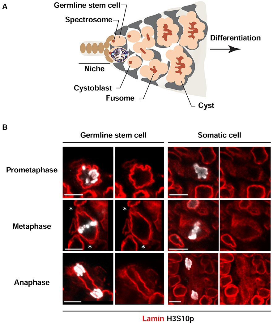
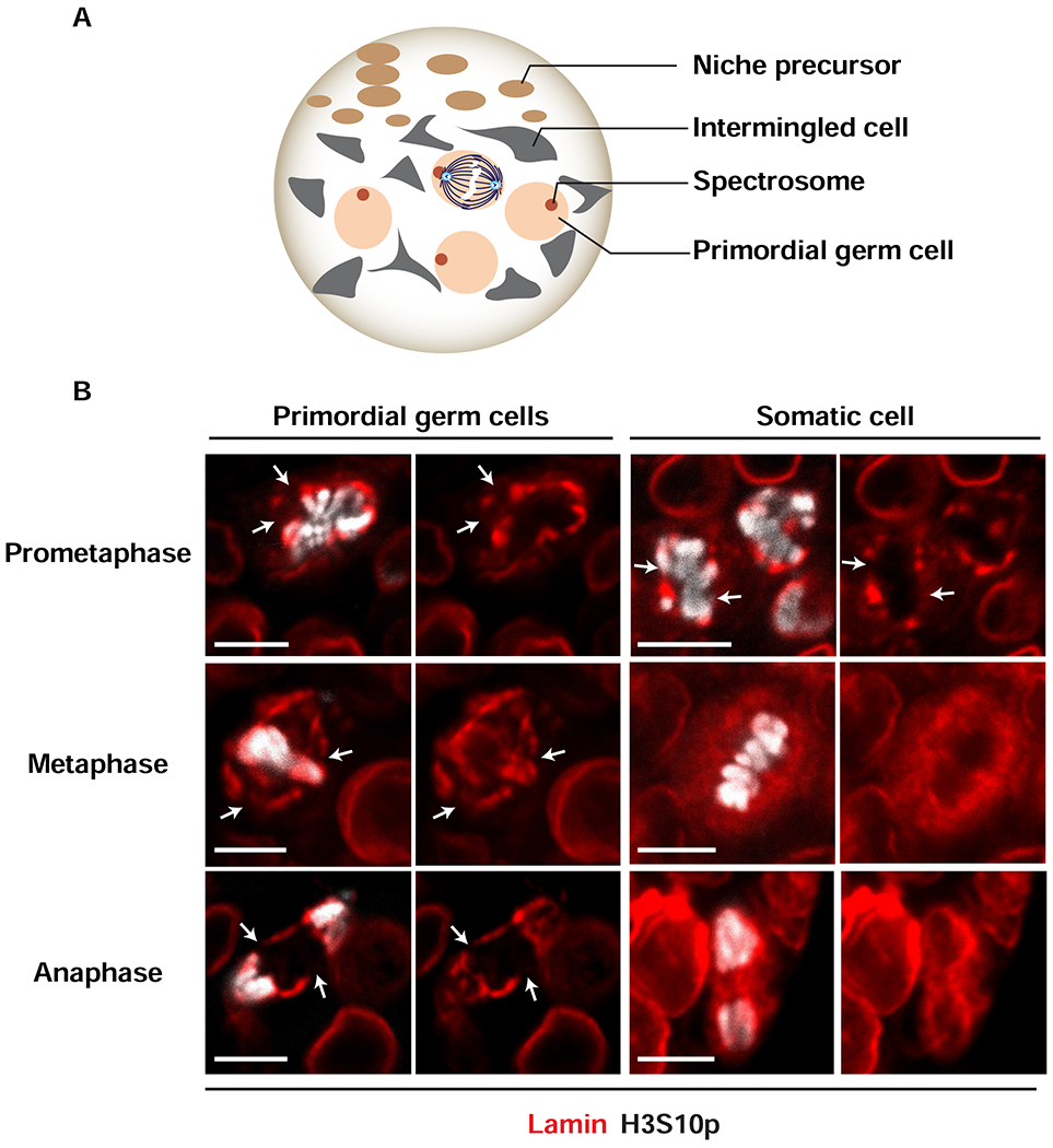
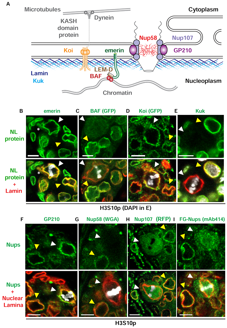
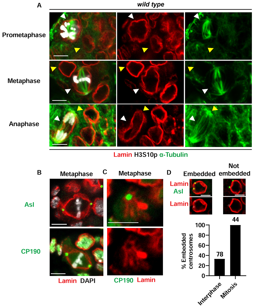
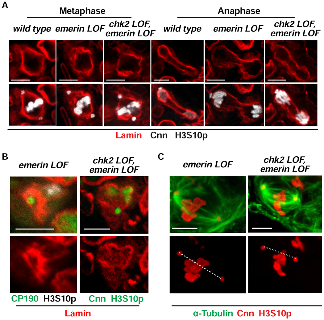
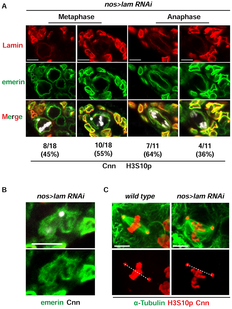
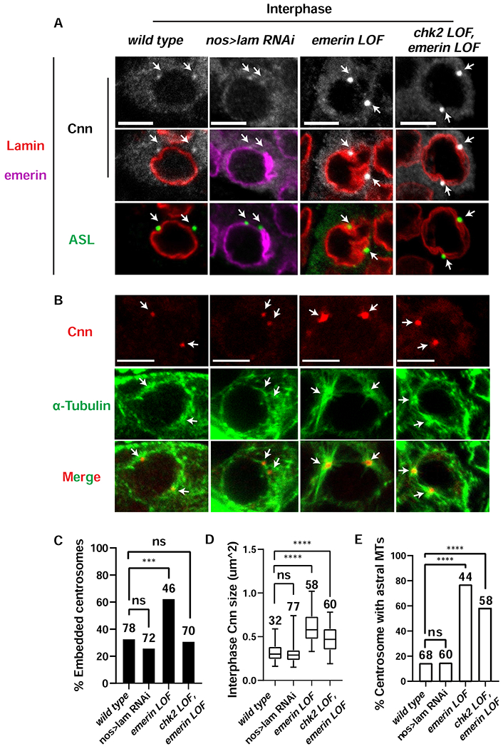
Stem cell homeostasis requires nuclear lamina (NL) integrity. In Drosophila germ cells, compromised NL integrity activates the ataxia telangiectasia and Rad3-related (ATR) and checkpoint kinase 2 (Chk2) checkpoint kinases, blocking germ cell differentiation and causing germline stem cell (GSC) loss. Checkpoint activation occurs upon loss of either the NL protein emerin or its partner barrier-to-autointegration factor, two proteins required for nuclear reassembly at the end of mitosis. Here, we examined how mitosis contributes to NL structural defects linked to checkpoint activation. These analyses led to the unexpected discovery that wild-type female GSCs utilize a non-canonical mode of mitosis, one that retains a permeable but intact nuclear envelope and NL. We show that the interphase NL is remodeled during mitosis for insertion of centrosomes that nucleate the mitotic spindle within the confines of the nucleus. We show that depletion or loss of NL components causes mitotic defects, including compromised chromosome segregation associated with altered centrosome positioning and structure. Further, in emerin mutant GSCs, centrosomes remain embedded in the interphase NL. Notably, these embedded centrosomes carry large amounts of pericentriolar material and nucleate astral microtubules, revealing a role for emerin in the regulation of centrosome structure. Epistasis studies demonstrate that defects in centrosome structure are upstream of checkpoint activation, suggesting that these centrosome defects might trigger checkpoint activation and GSC loss. Connections between NL proteins and centrosome function have implications for mechanisms associated with NL dysfunction in other stem cell populations, including NL-associated diseases, such as laminopathies.

Keywords: Drosophila oogenesis; LEM-domain proteins; barrier-to-autointegration factor; centrosome; checkpoint kinase 2; emerin; germline stem cells; mitosis; nuclear lamina; otefin.


Conflict of interest statement

Declaration of interests The authors declare no competing interests.

Figures

Figure 1.
Figure 1.. Lamin-B does not disperse during mitoses of adult GSCs.
(A) Shown is a schematic of a germarium, the structure that houses the stem cell niche in the adult ovary. Somatic cells of the germarium include cells comprising the niche (brown) and lining the anterior of the germarium (grey). Each niche anchors two or three germline stem cells (GSCs, peach). Asymmetric GSC divisions result from anchoring one pole of the mitotic spindle to the anterior spectrosome (red), located at the niche-stem cell interface. Asymmetric GSC divisions produce one self-renewing daughter that remains at the niche and a second differentiating daughter, cystoblast (peach) that enters into synchronized interconnected mitotic divisions anchored to a branched spectrosome or fusome (branched red structure). (B) Shown are representative nuclear images of wild type GSCs and somatic mitotic cells in adult ovaries stained with antibodies against Vasa (not shown), Lamin-B (red) and H3S10p (white). Stages of mitosis are labeled on the left. Asterisks indicate locations of Lamin-B looped structures (centrosomes). Scale bars: 5 μm. Numbers of nuclei analyzed are summarized in the Table S1.
Figure 2.
Figure 2.. Cell-type specific structural changes in NL in mitotic cells in larval ovaries.
(A) Shown is a schematic of a mid-third instar larval ovary. At this stage, the ovary carries ~100 primordial germ cells (PGCs, peach) that are identified by a spherical spectrosome (red) and multiple types of somatic cells, including anterior niche precursor cells (brown) and intermingled cells (grey). Expansion of PGC numbers occurs by random orientation of symmetric mitotic divisions (center) that produce two daughter PGCs. (B) Shown are representative nuclear images from wild type PGCs and somatic cells that are undergoing mitosis in the mid-third instar larval ovary. Ovaries were stained with antibodies against Vasa to identify germ cells (not shown), Lamin-B (red) to identify the NL and H3S10p (white) to identify cells in mitosis (stages of mitosis are labeled on the left).White arrow indicate locations of breaks in the NL. Scale bars: 5 μm. Numbers of nuclei analyzed are summarized in Table S1.
Figure 3.
Figure 3.. The mitotic NL differs in composition from the interphase NL.
(A) Shown is a schematic of proteins found in interphase NE and NL. Outer and inner nuclear membranes are fused at nuclear pores, where NPCs are located. NPCs are anchored to the NE by a transmembrane ring complex that includes GP210 (purple). This complex is connected to the Nup107 complex (light purple) that forms the core scaffold of the NPC and associates with the central FG-repeat Nups that include Nup58 (red). The NL lies underneath the inner nuclear membrane. The NL contains two farnesylated proteins, Lamin-B (dark blue) and Kugelkern (Kuk, light blue). Other NL proteins include the LEM-D protein emerin (green) that interacts with BAF (red) and the SUN domain protein Klaroid (Koi, orange) that interacts with KASH domain proteins (grey) to form the LINC complex that connects to the cytoskeleton through motor proteins, such as dynein (grey). (B-E) Confocal images of wild type metaphase fGSC nuclei in ovaries of newly born females that were stained with antibodies against several NL proteins (green, top) and co-stained with Lamin-B (red, bottom) and H3S10p (B-D, white) or DAPI (E, white). White arrowheads point to metaphase fGSCs. Yellow arrowheads point to interphase cells. Asterisks indicate locations of the Lamin-B loop structures (centrosome). Scale bar: 5 μm. (F-I) Confocal images of wild type metaphase fGSC nuclei in ovaries of newly born females that were stained with antibodies against several NPC components (green, top), NL proteins (red, merge bottom) and H3S10p (white). The NL protein corresponds to emerin in the co-stain with GP210 (F) or Lamin-B in the co-stain with the other Nups (G-I). In Drosophila, Wheat Germ Agglutinin (WGA) staining primarily reflects the localization of one FG-Nup, Nup 58 [100]. White arrowheads point to metaphase fGSCs. Yellow arrowheads point to interphase cells. Scale bar: 5 μm. Numbers of nuclei examined for each staining are summarized in Table S1. See also Figures S1 and S2.
Figure 4.
Figure 4.. The mitotic spindle is nucleated from centrosomes embedded within the NL.
(A) Shown are confocal images of wild type GSC nuclei in ovaries that were stained with antibodies against α-Tubulin (Green) Lamin-B (red) and H3S10p (white). Mitotic stages are labeled on the left. White arrowheads point to metaphase fGSCs. Yellow arrowheads point to interphase cells. Scale bars: 5 μm. (B) Confocal images of wild type metaphase fGSC nuclei in ovaries that were co-stained with antibodies against centrosome components, Asl and CP190 (green), Lamin-B (red) and DAPI (white). Scale bars: 5 μm. (C) Shown is a confocal image focused on the centrosome region in a wild type fGSC mitotic nucleus in an ovary that was stained with antibodies against CP190 (green) and Lamin-B (red). Scale bars: 5 μm. (D) Top: Shown are confocal images of wild type interphase fGSCs stained with antibodies against Asl (green) and Lamin-B (red). Examples of fGSCs with embedded or not embedded centrosomes are shown. Embedding is defined by a greater than 50% overlap between the centrosome and the NL. Bottom: Bar graph showing the percentage of embedded centrosomes in wild type GSCs. Stages of the cell cycle are listed at the bottom and the number of GSCs scored is listed at the top. Numbers of nuclei examined for each staining in (A)-(C) are summarized in Table S1. See also Figure S2.
Figure 5.
Figure 5.. Loss of emerin disrupts mitotic NL structure and spindle formation.
(A) Representative confocal images of metaphase and anaphase fGSC nuclei from ovaries of the indicated genotype that were stained with antibodies against Lamin-B (red), H3S10p (white) and Cnn (white). (B) Shown are confocal images of mitotic nuclei focusing on centrosome regions in emerin and chk2, emerin mutant fGSCs in ovaries that were stained with antibodies against centrosomal proteins CP190 or Cnn (green), H3S10p (white on the left and green on the right) and Lamin-B (red). Scale bars: 5 μm. (C) Representative images of metaphase fGSC nuclei in emerin and chk2, emerin mutant ovaries that were stained with antibodies against α-Tubulin (green), Cnn (red) and H3S10p (red). The dashed line connects the centers of two centrosomes. Quantification of centrosome positioning can be found in Figure S3B. Scale bar: 5 μm. Numbers of nuclei examined for each staining are summarized in Table S1. See also Figures S3 and S4.
Figure 6.
Figure 6.. Lamin-B is required for fGSC mitosis.
(A) Representative confocal images of metaphase and anaphase fGSC nuclei found in nos>lam RNAi ovaries that were stained with antibodies against Lamin-B (red), emerin (green), H3S10p (white) and Cnn (white). The number and percentage of nuclei found in each category are denoted below each image. (B) Shown is a confocal image of a mitotic nos>lam RNAi nucleus, focused on the centrosome region from ovaries stained with antibodies against emerin (green) and Cnn (white). Scale bars: 5 μm. (C) Representative images of metaphase fGSC nuclei isolated from wild type and nos>lamin RNAi ovaries that were stained with antibodies against α-Tubulin (green), Cnn (red) and H3S10p (red). The dashed line connects the centers of the centrosomes. Quantification of centrosome positioning can be found in Figure S3A. Scale bar: 5 μm. Numbers of nuclei examined for each staining are summarized in Table S1. See also Figure S3.
Figure 7.
Figure 7.. PCM retention in emerin mutants correlates with checkpoint activation.
(A) Confocal images of interphase fGSC nuclei in ovaries that were stained with antibodies against Lamin-B (red), emerin (purple), the centriolar marker Asterless (Asl; green) and the PCM marker Cnn (white). Genotypes are indicated along the top. Scale bars represent 5 μm. Arrows point to the location of centrosomes. (B) Confocal images of interphase fGSCs in ovaries that were stained with antibodies against H3S10p (not shown), Cnn (red) and α-Tubulin (green). Genotypes are the same as in (A). Scale bars represent 5 μm. Arrows point to the location of centrosomes. (C) Shown is a bar graph comparing the percentage of NL embedded centrosomes in interphase fGSCs of the indicated genotype. Embedded centrosomes were defined as centrosomes that showed at least 50% overlap with the NL staining. The number of centrosomes assessed is noted above each bar. Asterisks indicate significance [Two proportion Z-test (ns: not significant, *** < 0.001)]. (D) Shown are box plots of the size of centrosomal Cnn in interphase fGSCs of the indicated genotype. For each box plot, the box represents the 25th to 75th percentile interval, the line represents the median and the whiskers represent the 5th to 95th percentile interval and non-outlier range. The number of centrosomes analyzed is noted above each box. Asterisks indicate significance [Mann-Whitney U-test, **** <0.0001, ns: not significant]. (E) Shown is a bar graph comparing the percentage of interphase centrosomes that nucleate aster microtubules (MT). The number of centrosomes quantified is noted above each bar. Asterisks indicate significance using the two proportion Z-test (ns: not significant; ****<0.0001). See also Figure S5 and Table S1.

Similar articles

Cited by

References

    1. Gruenbaum Y, Margalit A, Goldman RD, Shumaker DK, and Wilson KL (2005). The nuclear lamina comes of age. Nat Rev Mol Cell Biol 6, 21–31. - PubMed
    1. Baxley RM, Soshnev AA, Koryakov DE, Zhimulev IF, and Geyer PK (2011). The role of the Suppressor of Hairy-wing insulator protein in Drosophila oogenesis. Dev Biol 356, 398–410. - PMC - PubMed
    1. Goldman RD, Gruenbaum Y, Moir RD, Shumaker DK, and Spann TP (2002). Nuclear lamins: building blocks of nuclear architecture. Genes Dev 16, 533–547. - PubMed
    1. Gonzalo S (2014). DNA Damage and Lamins. Cancer Biology and the Nuclear Envelope: Recent Advances May Elucidate Past Paradoxes 773, 377–399. - PMC - PubMed
    1. Burke B, and Stewart CL (2014). Functional architecture of the cell’s nucleus in development, aging, and disease. Curr Top Dev Biol 109, 1–52. - PubMed

Publication types