

Diagnostic and management challenges for MDM2-, CDK4-negative fatty tumors of the retroperitoneum
- PMID: 33550433
- PMCID: PMC11802130
- DOI: 10.1007/s00432-021-03512-x
Diagnostic and management challenges for MDM2-, CDK4-negative fatty tumors of the retroperitoneum
Abstract
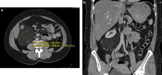
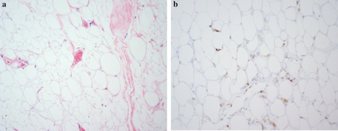
Background: Neoplasms of the retroperitoneum that contain a major fat component may represent either benign entities, such as lipomas or angiomyolipomas, or malignancy such as liposarcoma. Distinguishing these diagnoses has important implications for management. While liposarcomas often stain positively for MDM2 and CDK4 proteins, absence of these markers can lead to diagnostic and management challenges.
Methods: We examined three cases in our institution of fat-containing masses of the retroperitoneum that lacked MDM2 and CDK4 markers to highlight the challenges in diagnosing and managing these cases. A thorough review of the literature examining radiologic and histologic features that can be used to determine that diagnosis was conducted and summarized.
Results: The three cases we present represent the three main diagnostic entities that can be found in among fatty tumors of the retroperitoneum: lipoma, angiomyolipoma, and liposarcoma. While radiologic features and analysis of histology helped to inform management, these cases in conjunction with the literature also illustrate the limitations of the diagnostic work up and importance also factoring the biologic behavior of the tumor in its management.
Conclusion: Fat-containing tumors of the retroperitoneum that do not stain for MDM2 or CDK4 can pose a diagnostic challenge. Assessing radiologic and pathologic features in conjunction with the biologic behavior of these tumors should inform their management.
Keywords: Angiomyolipoma; CDK4; MDM2; Retroperitoneal fatty tumor; Well-differentiated liposarcoma.
Conflict of interest statement
The authors declare that they have no conflict of interest.
Figures


Similar articles
-
Can HMGI-C be used as an aid with MDM2 and CDK4 to differentiate liposarcoma subtypes from their mimics?J Cancer Res Clin Oncol. 2013 Jun;139(6):1073-81. doi: 10.1007/s00432-013-1420-6. Epub 2013 Mar 26. J Cancer Res Clin Oncol. 2013. PMID: 23529275 Free PMC article.
-
IMP3, CDK4, MDM2 and β-catenin expression in Enchondroma and Central Chondrosarcoma: Diagnostic and prognostic utility.Clinics (Sao Paulo). 2024 Oct 4;79:100483. doi: 10.1016/j.clinsp.2024.100483. eCollection 2024. Clinics (Sao Paulo). 2024. PMID: 39368400 Free PMC article.
-
Signs and symptoms to determine if a patient presenting in primary care or hospital outpatient settings has COVID-19.Cochrane Database Syst Rev. 2022 May 20;5(5):CD013665. doi: 10.1002/14651858.CD013665.pub3. Cochrane Database Syst Rev. 2022. PMID: 35593186 Free PMC article.
-
Behavioral interventions to reduce risk for sexual transmission of HIV among men who have sex with men.Cochrane Database Syst Rev. 2008 Jul 16;(3):CD001230. doi: 10.1002/14651858.CD001230.pub2. Cochrane Database Syst Rev. 2008. PMID: 18646068
-
Can a Liquid Biopsy Detect Circulating Tumor DNA With Low-passage Whole-genome Sequencing in Patients With a Sarcoma? A Pilot Evaluation.Clin Orthop Relat Res. 2025 Jan 1;483(1):39-48. doi: 10.1097/CORR.0000000000003161. Epub 2024 Jun 21. Clin Orthop Relat Res. 2025. PMID: 38905450
Cited by
-
A case report and literature review on a large MDM2 negative retroperitoneal/psoas muscle well-differentiated liposarcoma mimicking intramuscular myxoma.Radiol Case Rep. 2025 Jun 26;20(9):4661-4668. doi: 10.1016/j.radcr.2025.05.094. eCollection 2025 Sep. Radiol Case Rep. 2025. PMID: 40677885 Free PMC article.
-
Retroperitoneal lipoma and bilateral renal cell carcinoma in a rare co-existence.Int J Surg Case Rep. 2022 Oct;99:107718. doi: 10.1016/j.ijscr.2022.107718. Epub 2022 Oct 3. Int J Surg Case Rep. 2022. PMID: 36261952 Free PMC article.
References
-
- Arbiser ZK, Folpe AL, Weiss SW (2001) Consultative (expert) second opinions in soft tissue pathology. Analysis of problem-prone diagnostic situations. Am J ClinPathol 116(4):473–476 - PubMed
-
- Bonvalot S, Rivoire M, Castaing M et al (2009) Primary retroperitoneal sarcomas: a multivariate analysis of surgical factors associated with local control. J ClinOncol 27(1):31–37 - PubMed
-
- Dal Cin P, Kools P, Sciot R et al (1993) Cytogenetic and fluorescence in situ hybridization investigation of ring chromosomes characterizing a specific pathologic subgroup of adipose tissue tumors. Cancer Genet Cytogenet 68(2):85–90 - PubMed
Publication types
MeSH terms
Substances
LinkOut - more resources
Full Text Sources
Other Literature Sources
Research Materials
