Case Reports
. 2021 Jul 1;60(13):2119-2123.
doi: 10.2169/internalmedicine.6572-20. Epub 2021 Feb 8.

Composite Epstein-Barr Virus-associated T-lymphoblastic and Peripheral T-cell Lymphomas: A Clonal Study

Affiliations
Case Reports

Composite Epstein-Barr Virus-associated T-lymphoblastic and Peripheral T-cell Lymphomas: A Clonal Study

Hiroki Hosoi et al. Intern Med. .

Abstract

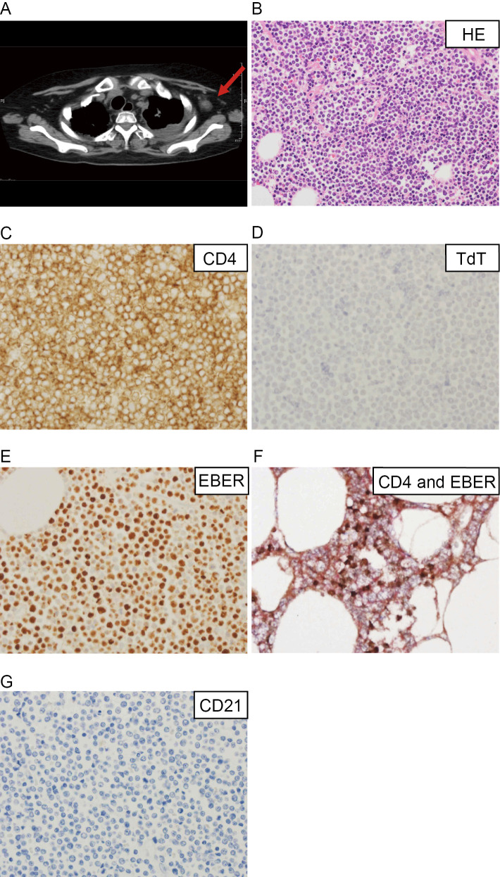
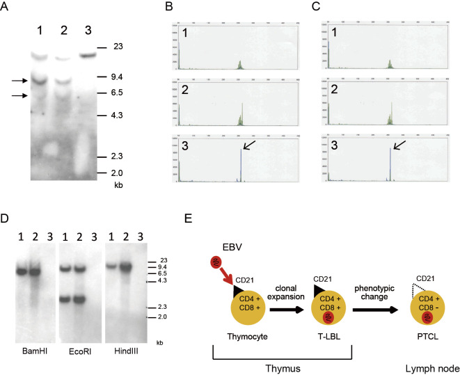
A 30-year-old woman was diagnosed with T-lymphoblastic lymphoma (T-LBL) that harbored a clonal Epstein-Barr virus (EBV) genome. At relapse, axillary lymph node adenopathy, which was diagnosed as peripheral T-cell lymphoma, not otherwise specified (PTCL-NOS), was detected. Southern blot analyses of the T-cell receptor and EBV genome revealed that the T-LBL and PTCL-NOS were clonally identical. We previously showed that CD21 acted as an entry molecule that allowed EBV into the patient's T-LBL cells. Interestingly, the PTCL-NOS cells lacked CD21 expression. Our case suggests that EBV might infect immature CD21-positive T-cells, and CD21-negative PTCL-NOS might subsequently arise through phenotypic changes.

Keywords: CD21; Epstein-Barr virus; T-lymphoblastic lymphoma; composite lymphoma; peripheral T-cell lymphoma.


Conflict of interest statement

The authors state that they have no Conflict of Interest (COI).

Figures

Figure 1.
Figure 1.
Computed tomography (CT) and histopathological findings of the patient at relapse. A: CT image of the patient obtained at relapse. The red arrow indicates the left axillary lymph nodes resected during the postmortem examination. B-G: Histopathological findings of the peripheral T-cell lymphoma, not otherwise specified (PTCL-NOS), after it was resected from the axillary lymph nodes. B: Hematoxylin and Eosin staining (×400); C, D: Immunohistochemistry of CD4 (C) and TdT (D) (×600); E: In situ hybridization of Epstein-Barr virus (EBV)-encoded mRNA (×600). F: Double immunohistochemical staining of CD4 and EBER. The CD4- and EBER-positive cells were stained red and brown, respectively (×600). G: Immunohistochemistry of CD21 (×600).
Figure 2.
Figure 2.
Genomic analyses of the T-cell receptor (TCR) and EBV. A: Southern blot analyses using a Cβ2 probe. The arrows indicate rearranged bands. DNA was digested with BamHI. Lane 1: peripheral lymph nodes, lane 2: pleural effusion, lane 3: MD901 (the negative control). B, C: TCR gene rearrangement of Vβ/Jβ1/Jβ2 in cells from the peripheral lymph nodes (B) or pleural effusion (C). The polymerase chain reaction was conducted using 23 Vβ primers and 9 Jβ primers, as described previously (9). The arrows indicate the clonal peaks. The same clonal peaks were seen in (B) and (C), indicating that these cells were identical. 1: negative control, 2: positive control, 3: patient’s sample. D: Southern blot analyses of the EBV genome. The EBV1 and EBV2 fragments were used as probes. DNA was digested with BamHI, EcoRI, and HindIII. Lane 1: peripheral lymph nodes, lane 2: pleural effusion, lane 3: MD901 (the negative control). E: Schematic diagram showing a putative mechanism that might have caused the EBV-induced T-cell malignancies seen in this patient. EBV-infected, CD4-positive, CD8-positive immature T-cells, which also expressed CD21, were present in the patient’s thymus. The cells developed into T-lymphoblastic lymphoma (T-LBL). After treatment, including allogeneic stem cell transplantation, CD4-positive, CD8-negative PTCL developed through phenotypic changes. The PTCL cells were negative for CD21, which was the molecule responsible for the entry of EBV into the T-LBL cells in this patient, but EBV had already infected the PTCL cells.

Similar articles

References

    1. Young LS, Yap LF, Murray PG. Epstein-Barr virus: more than 50 years old and still providing surprises. Nat Rev Cancer 16: 789-802, 2016. - PubMed
    1. Fox CP, Shannon-Lowe C, Rowe M. Deciphering the role of Epstein-Barr virus in the pathogenesis of T and NK cell lymphoproliferations. Herpesviridae 2: 8, 2011. - PMC - PubMed
    1. Hutt-Fletcher LM. Epstein-Barr virus entry. J Virol 81: 7825-7832, 2007. - PMC - PubMed
    1. Braun M, Melchers I, Peter HH, Illges H. Human B and T lymphocytes have similar amounts of CD21 mRNA, but differ in surface expression of the CD21 glycoprotein. Int Immunol 10: 1197-1202, 1998. - PubMed
    1. Castillo JJ, Reagan JL, Bishop KD, Apor E. Viral lymphomagenesis: from pathophysiology to the rationale for novel therapies. Br J Haematol 165: 300-315, 2014. - PubMed

Publication types