. 2021 Jan 21:11:614928.
doi: 10.3389/fphar.2020.614928. eCollection 2020.

A Putative Serine Protease is Required to Initiate the RIPK3-MLKL-Mediated Necroptotic Death Pathway in Neutrophils

Affiliations

A Putative Serine Protease is Required to Initiate the RIPK3-MLKL-Mediated Necroptotic Death Pathway in Neutrophils

Xiaoliang Wang et al. Front Pharmacol. .

Abstract

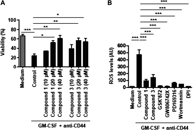
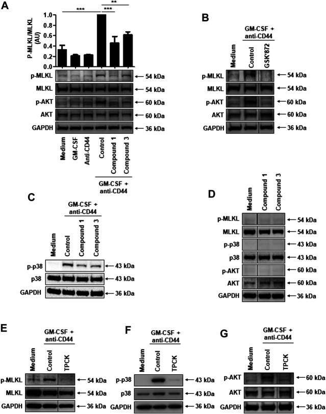
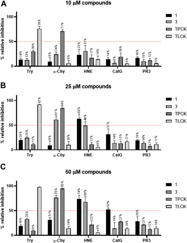
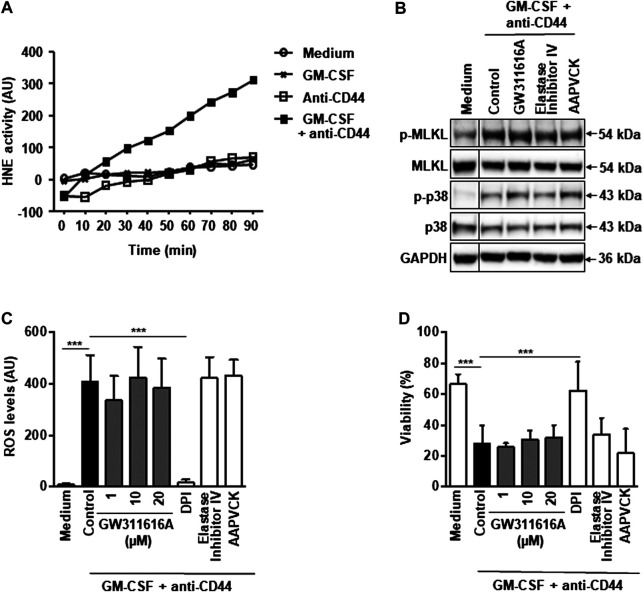
Adhesion receptors, such as CD44, have been shown to activate receptor interacting protein kinase-3 (RIPK3)-mixed lineage kinase-like (MLKL) signaling, leading to a non-apoptotic cell death in human granulocyte/macrophage colony-stimulating factor (GM-CSF) - primed neutrophils. The signaling events of this necroptotic pathway, however, remain to be investigated. In the present study, we report the design, synthesis, and characterization of a series of novel serine protease inhibitors. Two of these inhibitors, compounds 1 and 3, were able to block CD44-triggered necroptosis in GM-CSF-primed neutrophils. Both inhibitors prevented the activation of MLKL, p38 mitogen-activated protein kinase (MAPK) and phosphatidylinositol 3'-kinase (PI3K), hence blocking the increased levels of reactive oxygen species (ROS) required for cell death. Although compounds one and three partially inhibited isolated human neutrophil elastase (HNE) activity, we obtained no pharmacological evidence that HNE is involved in the initiation of this death pathway within a cellular context. Interestingly, neither serine protease inhibitor had any effect on FAS receptor-mediated apoptosis. Taken together, these results suggest that a serine protease is involved in non-apoptotic CD44-triggered RIPK3-MLKL-dependent neutrophil cell death, but not FAS receptor-mediated caspase-dependent apoptosis. Thus, a pharmacological block on serine proteases might be beneficial for preventing exacerbation of disease in neutrophilic inflammatory responses.

Keywords: necroptosis; neutrophil; serine protease; signal transduction; small molecule inhibitor.


Conflict of interest statement

The authors declare that the research was conducted in the absence of any commercial or financial relationships that could be construed as a potential conflict of interest.

Figures

FIGURE 1
FIGURE 1
CD44-induced ROS production and subsequent necroptosis in GM-CSF-primed neutrophils require serine protease activity. (A) Concentration-dependent effect of serine protease inhibitors, compounds 1 or 3, on CD44-triggered necroptosis in GM-CSF-primed neutrophils. GM-CSF-primed neutrophils were precultured in the presence and absence of compound 1 (10, 20, or 40 μM) or compound 3 (10, 20, or 40 μM), and subsequently stimulated with anti-CD44 mAb for 24 h. Cell death was assessed by uptake of 25 μM ethidium bromide with flow cytometric analysis. (B) DHR oxidation assay. GM-CSF-primed neutrophils were precultured in the presence and absence of the serine protease inhibitor compounds 1 (40 μM) or 3 (20 μM), the RIPK3 inhibitors GSK′872 (10 μM), the MLKL inhibitors GW806742X (5 μM), the p38 inhibitor PD169316 (10 μM), the PI3K inhibitor wortmannin (100 nM), or the NADPH oxidase inhibitor DPI (1 μM) and subsequently stimulated with anti-CD44 mAb for 15 min before measured by flow cytometry. All values are means ± SEM of at least 3 independent experiments. *p < 0.05, **p < 0.01, ***p < 0.001.
FIGURE 2
FIGURE 2
Serine protease activity is required for RIPK3-dependent MLKL phosphorylation, and subsequent p38 MAPK and PI3K activation in CD44-induced necroptosis in GM-CSF-primed neutrophils. (A–C and E–G) Immunoblotting. Neutrophils were cultured as indicated and subsequently stimulated with anti-CD44 mAb for 5 min. (D) Immunoblotting. Neutrophils were cultured in the presence and absence of compound 1 (40 μM) or compound 3 (20 μM) for 1 h. Cell lysates were analyzed by immunoblotting for phosphorylated MLKL, phosphorylated AKT, and phosphorylated p38 MAPK. MLKL, AKT, p38, and GAPDH expression levels were analyzed as loading controls. Representative immunoblots are shown (n ≥ 3). (A) Upper panel: p-MLKL protein expression levels were quantified relative to the control condition of MLKL. Data are from 3 independent experiments. **p < 0.01, ***p < 0.001.
FIGURE 3
FIGURE 3
Comparative activity of compounds against five different serine proteases. Compound 1, 3, TPCK, and TLCK were incubated with trypsin (Try), α-chymotrypsin (α-Chy), human neutrophil elastase (HNE), cathepsin G (CatG), and proteinase 3 (PR3) at 37 °C for 30 min. Subsequently, reactions were started by adding an enzyme-specific substrate. Substrate hydrolysis was monitored in a kinetic mode for 60 min. The initial rates of reactions were used to calculate the % relative inhibition of the enzymes. Results are presented as mean ± SEM of 3 independent experiments, run in duplicate. (A) Relative inhibition (%) of serine proteases for 10 µM compound 1, 3, TPCK, and TLCK. (B) Relative inhibition (%) of serine proteases for 25 µM compound 1, 3, TPCK, and TLCK. (C) Relative inhibition (%) of serine proteases for 50 µM compound 1, 3, TPCK, and TLCK.
FIGURE 4
FIGURE 4
Pharmacological inactivation of HNE does not block CD44 death signaling in GM-CSF-primed neutrophils. (A) Enzymatic assay. Supernatant HNE activity can be measured upon CD44 activation in GM-CSF-primed neutrophils. Data are representative of 3 independent experiments. (B) Immunoblotting. Neutrophils were cultured as indicated and subsequently stimulated with anti-CD44 mAb for 5 min. Preincubation with GW311616A (10 μM), AAPVCK (10 μM), or elastase inhibitor IV (10 μM) was performed for 30 min. Cell lysates were analyzed by immunoblotting for phosphorylated MLKL and phosphorylated p38 MAPK. MLKL, p38 and GAPDH expression levels were analyzed as loading controls. (C) DHR oxidation assay. GM-CSF-primed neutrophils were precultured in the presence and absence of GW311616A (1, 10, and 20 μM), AAPVCK (10 μM), or elastase inhibitor IV (10 μM), and subsequently stimulated with anti-CD44 mAb for 15 min before measured by flow cytometry. DPI (1 μM) served as a control. (D) Viability assay. GM-CSF-primed neutrophils were precultured in the presence and absence of GW311616A (1, 10, and 20 μM), AAPVCK (10 μM), or elastase inhibitor IV (10 μM) for 30 min and subsequently stimulated with anti-CD44 mAb for 24 h. Cell death was assessed by uptake of 25 μM ethidium bromide with flow cytometric analysis. All values are means ± SEM of at least 3 independent experiments. ***p < 0.001.

References

    1. Amini P., Stojkov D., Wang X., Wicki S., Kaufmann T., Wong W. W., et al. (2016). NET formation can occur independently of RIPK3 and MLKL signaling. Eur. J. Immunol. 46, 178–184. 10.1002/eji.201545615 - DOI - PMC - PubMed
    1. Amini P., Stojkov D., Felser A., Jackson C. B., Courage C., Schaller A., et al. (2018). Neutrophil extracellular trap formation requires OPA1-dependent glycolytic ATP production. Nat. Commun. 9, 2958 10.1038/s41467-018-05387-y - DOI - PMC - PubMed
    1. Barbosa L. A., Fiuza P. P., Borges L. J., Rolim F. A., Andrade M. B., Luz N. F., et al. (2018). RIPK1-RIPK3-MLKL-associated necroptosis drives Leishmania infantum killing in neutrophils. Front. Immunol. 9, 1818 10.3389/fimmu.2018.01818 - DOI - PMC - PubMed
    1. Baumann M., Pham C. T., Benarafa C. (2013). SerpinB1 is critical for neutrophil survival through cell-autonomous inhibition of cathepsin G. Blood. 121, 3900–3906. 10.1182/blood-2012-09-455022 - DOI - PMC - PubMed
    1. Benarafa C., Simon H. U. (2017). Role of granule proteases in the life and death of neutrophils. Biochem. Biophys. Res. Commun. 482, 473–481. 10.1016/j.bbrc.2016.11.086 - DOI - PubMed