. 2021 Jan 20:11:619629.
doi: 10.3389/fpsyt.2020.619629. eCollection 2020.

Brain Age Prediction With Morphological Features Using Deep Neural Networks: Results From Predictive Analytic Competition 2019

Affiliations

Brain Age Prediction With Morphological Features Using Deep Neural Networks: Results From Predictive Analytic Competition 2019

Angela Lombardi et al. Front Psychiatry. .

Abstract

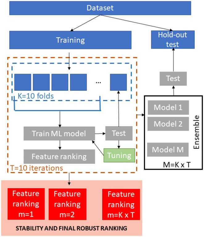
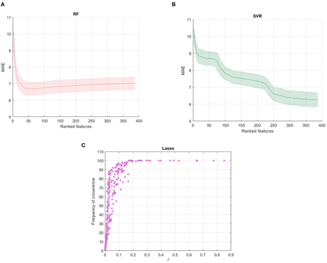
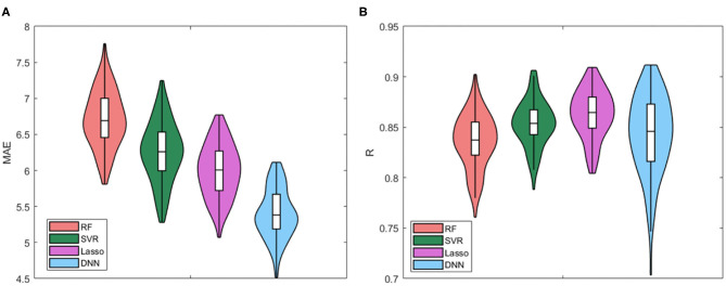
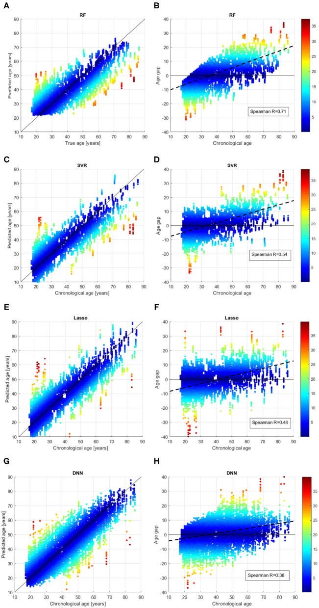
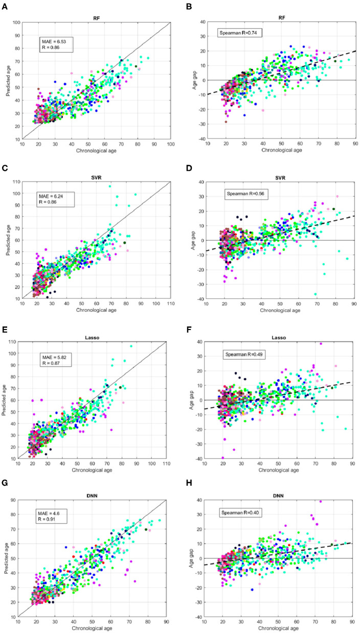
Morphological changes in the brain over the lifespan have been successfully described by using structural magnetic resonance imaging (MRI) in conjunction with machine learning (ML) algorithms. International challenges and scientific initiatives to share open access imaging datasets also contributed significantly to the advance in brain structure characterization and brain age prediction methods. In this work, we present the results of the predictive model based on deep neural networks (DNN) proposed during the Predictive Analytic Competition 2019 for brain age prediction of 2638 healthy individuals. We used FreeSurfer software to extract some morphological descriptors from the raw MRI scans of the subjects collected from 17 sites. We compared the proposed DNN architecture with other ML algorithms commonly used in the literature (RF, SVR, Lasso). Our results highlight that the DNN models achieved the best performance with MAE = 4.6 on the hold-out test, outperforming the other ML strategies. We also propose a complete ML framework to perform a robust statistical evaluation of feature importance for the clinical interpretability of the results.

Keywords: FreeSurfer; MRI; aging biomarker; brain aging; deep neural networks; machine learning; morphological features.


Conflict of interest statement

The authors declare that the research was conducted in the absence of any commercial or financial relationships that could be construed as a potential conflict of interest.

Figures

Figure 1
Figure 1
Schematic overview of the ML framework.
Figure 2
Figure 2
Shadow performance curves with the average MAE achieved by all the stepwise models in cross-validation with the standard errors for (A) RF models and (B) SVR models; (C) averaged weights vs. frequency of occurrence of the features across all the validation rounds resulting from Lasso algorithm.
Figure 3
Figure 3
Violin plots of the distributions of (A) MAE values and (B) R values for the four models.
Figure 4
Figure 4
Results of brain age prediction for the training set in cross-validation rounds for (A) the RF model, (C) the SVR model, (E) the Lasso model, (G) the DNN model; results of age gap (Δ) for the training set in cross-validation rounds for (B) the RF model, (D) the SVR model, (F) the Lasso model, (H) the DNN model.
Figure 5
Figure 5
Results of brain age prediction for the independent test set for (A) the RF model, (C) the SVR model, (E) the Lasso model, (G) the DNN model; results of age gap (Δ) for the independent test set for (B) the RF model, (D) the SVR model, (F) the Lasso model, (H) the DNN model. Each color indicates a specific site.
Figure 6
Figure 6
Mean values and standard errors of MAE resulting from the independent hold out test set grouped by the different sites for the four models.
Figure 7
Figure 7
(A) Training sample size reported for bins of 5 years; (B) ensemble variability within the test set quantified as the standard deviation of the prediction error within the ensemble of models for each ML algorithm.
Figure 8
Figure 8
Overlapping between the top (A) 10 features; (B) 20 features; (C) 30 features of each couple of models.

References

    1. Cole JH, Franke K. Predicting age using neuroimaging: innovative brain ageing biomarkers. Trends Neurosci. (2017) 40:681–90. 10.1016/j.tins.2017.10.001 - DOI - PubMed
    1. Madan CR, Kensinger EA. Predicting age from cortical structure across the lifespan. Eur J Neurosci. (2018) 47:399–416. 10.1111/ejn.13835 - DOI - PMC - PubMed
    1. Aycheh HM, Seong JK, Shin JH, Na DL, Kang B, Seo SW, et al. Biological brain age prediction using cortical thickness data: a large scale cohort study. Front Aging Neurosci. (2018) 10:252. 10.3389/fnagi.2018.00252 - DOI - PMC - PubMed
    1. Erus G, Battapady H, Satterthwaite TD, Hakonarson H, Gur RE, Davatzikos C, et al. Imaging patterns of brain development and their relationship to cognition. Cereb Cortex. (2015) 25:1676–84. 10.1093/cercor/bht425 - DOI - PMC - PubMed
    1. Han LK, Dinga R, Hahn T, Ching CR, Eyler LT, Aftanas L, et al. Brain aging in major depressive disorder: results from the ENIGMA Major Depressive Disorder working group. Mol Psychiatry. (2020) 1–16. 10.1038/s41380-020-0754-0 - DOI - PMC - PubMed

LinkOut - more resources