. 2021 Jan 21:11:601921.
doi: 10.3389/fmicb.2020.601921. eCollection 2020.

Functional Insights From KpfR, a New Transcriptional Regulator of Fimbrial Expression That Is Crucial for Klebsiella pneumoniae Pathogenicity

Affiliations

Functional Insights From KpfR, a New Transcriptional Regulator of Fimbrial Expression That Is Crucial for Klebsiella pneumoniae Pathogenicity

Ana Érika Inácio Gomes et al. Front Microbiol. .

Abstract

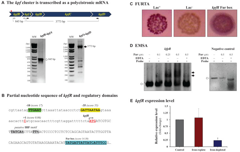
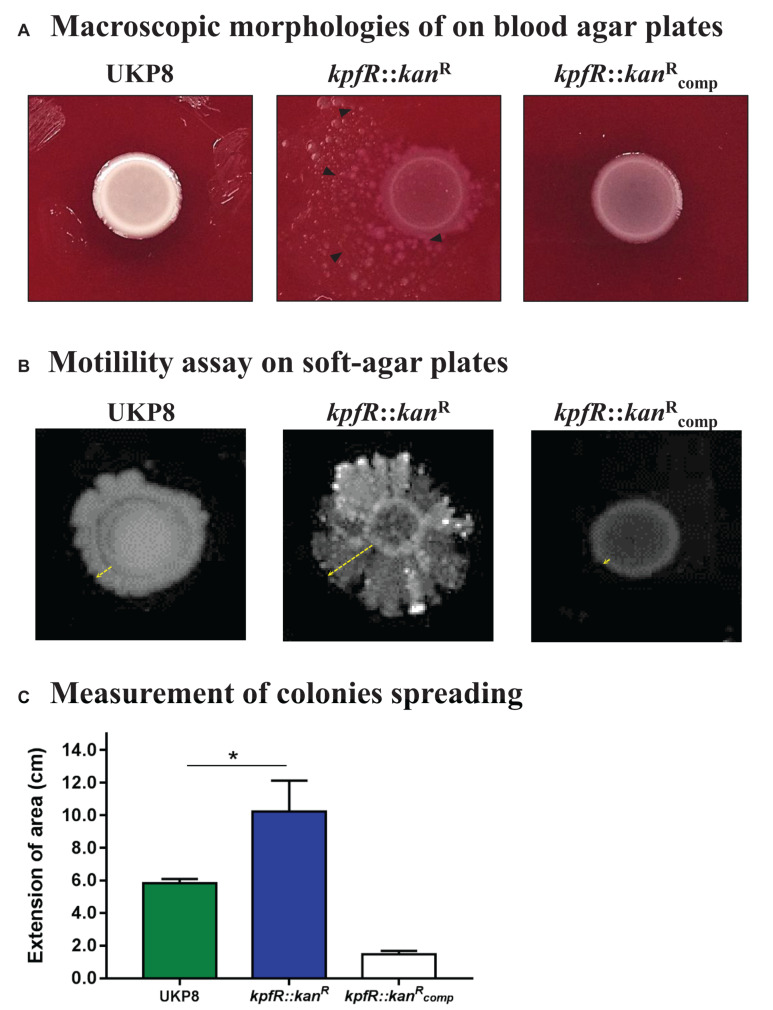
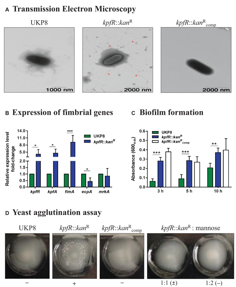
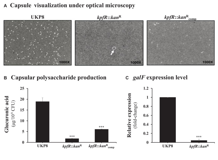
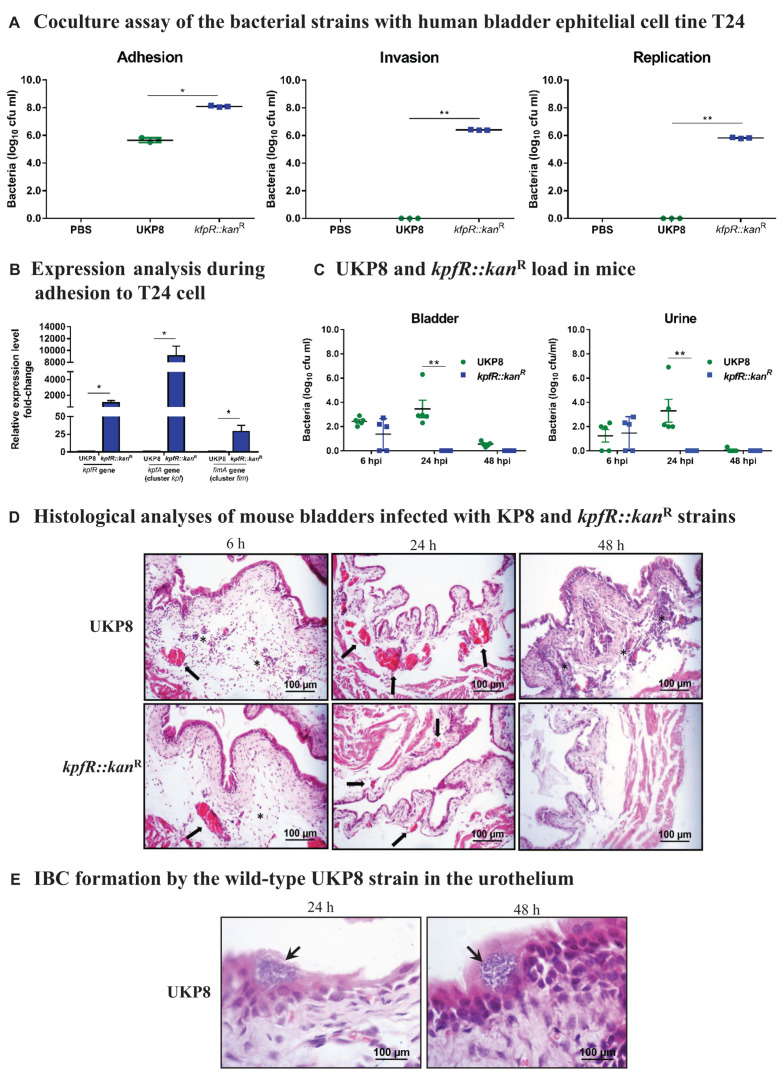
Although originally known as an opportunistic pathogen, Klebsiella pneumoniae has been considered a worldwide health threat nowadays due to the emergence of hypervirulent and antibiotic-resistant strains capable of causing severe infections not only on immunocompromised patients but also on healthy individuals. Fimbriae is an essential virulence factor for K. pneumoniae, especially in urinary tract infections (UTIs), because it allows the pathogen to adhere and invade urothelial cells and to form biofilms on biotic and abiotic surfaces. The importance of fimbriae for K. pneumoniae pathogenicity is highlighted by the large number of fimbrial gene clusters on the bacterium genome, which requires a coordinated and finely adjusted system to control the synthesis of these structures. In this work, we describe KpfR as a new transcriptional repressor of fimbrial expression in K. pneumoniae and discuss its role in the bacterium pathogenicity. K. pneumoniae with disrupted kpfR gene exhibited a hyperfimbriated phenotype with enhanced biofilm formation and greater adhesion to and replication within epithelial host cells. Nonetheless, the mutant strain was attenuated for colonization of the bladder in a murine model of urinary tract infection. These results indicate that KpfR is an important transcriptional repressor that, by negatively controlling the expression of fimbriae, prevents K. pneumoniae from having a hyperfimbriated phenotype and from being recognized and eliminated by the host immune system.

Keywords: Klebsiella pneumonia; adherence; biofilms; coculture; fimbriae; host-microbe interactions; transcriptional regulation; urinary tract infection.


Conflict of interest statement

The authors declare that the research was conducted in the absence of any commercial or financial relationships that could be construed as a potential conflict of interest.

Figures

FIGURE 1
FIGURE 1
The kpf gene cluster encoding type 1-like fimbriae is regulated by Fur. (A) kpf cluster comprises kpfR, kpfA, kpfB, kpfC, and kpfD genes and is transcribed as a polycistronic mRNA. cDNA synthesized from total RNA of K. pneumoniae was used in PCR reactions using the primer pairs represented in the scheme. The resulting amplicons were analyzed by agarose gel electrophoresis and confirmed the predicted sizes of 847 and 3,773 base pairs (bp). The red spot on the scheme indicates the putative Fur box sequence identified on kpf cluster. (B) Partial sequence of kpfR showing the initial codon (ATG, double underlined) and the Fur-binding sequence located inside the coding region of kpfR (highlighted in blue). Also indicated on the promoter region of the cluster are the –35 (in green) and –10 (in yellow) domains of the housekeeping Sigma factor 70, the predicted transcription initiation site at position + 1 (cytosine in red), and a putative IHF-binding consensus sequence (in gray; lowercase nucleotides are not identical to the consensus sequence). (C) Fur Titration Assay (FURTA) validated the Fur box on kpfR of K. pneumoniae, as indicated by the E. coli H1717 red colonies on MacConkey plates, similar to the FURTA-positive control (Lac+). (D) DNA Electrophoretic Mobility Shift Assay (EMSA) confirms the direct interaction of the K. pneumoniae Fur protein on the putative Fur box found on kpfR. Fur-DNA probe complexes are formed (mobility shifts, indicated by closed arrowheads) as increased concentrations of purified His-Fur protein from K. pneumoniae were incubated with the probes (DNA fragment containing the Fur box of kpfR). Fur interaction depends on divalent cations, since the addition of EDTA chelator abolished the mobility shift of the probe (open arrowheads). No mobility shift is observed in the EMSA with the negative control (DNA probe without Fur box), indicated by open arrowheads). (E) RT-qPCR analyses showed that Fur represses the expression of the kpfR gene on K. pneumoniae cells cultured under iron-depleted condition when compared to the control condition (bacteria cultured in LB medium only). Since kpfR belongs to the kpf gene cluster, Fur modulates the expression of the entire cluster according to the availability of iron in the culture medium. *p ≤ 0.05.
FIGURE 2
FIGURE 2
Bacterial colonies of K. pneumoniae cells depleted of kpfR show altered macroscopic morphologies and rudimentary motility. (A) Colony morphology of wild-type (UKP8), mutant (kpfR::kanR), and complemented (kpfR::kancompR) strains were investigated on blood agar plates. The altered pattern on the mutant strain was fully restored in the complemented strain, which exhibited a pattern similar to the wild-type. (B,C) In soft agar plates, kpfR::kanR cells exhibited a pronounced asymmetric dispersion on the surface of the plates when compared to UKP8, resembling a sort of rudimentary motility. Dispersion was only partially restored by complementation. The greater dispersion of the mutant colonies than the wild-type is statistically significant. *p ≤ 0.05.
FIGURE 3
FIGURE 3
Lack of KpfR triggers the expression of type 1-like fimbriae, increases biofilm formation and yeast cells agglutination, and reduces production of capsule polysaccharide. (A) TEM analyses revealed kpfR::kanR mutant cells covered with numerous fimbriae-like appendages that were absent in wild-type and complemented kpfR::kancompR cells. The images are representative of independent experiments. (B) This hyperfimbriated phenotype is corroborated by the fimbrial genes expression analyses. The ecpA gene, from ecp gene cluster, is downregulated, while fimA (fim gene cluster of type 1 fimbriae) and kpfR and kpfA genes (kpf gene cluster of type 1-like fimbriae) are up-regulated in the kpfR::kanR mutant cells. *p ≤ 0.05; **p ≤ 0.01; ***p ≤ 0.005. (C,D) Because of the hyperfimbriated phenotype, the mutant strain forms more biofilm than the wild-type and agglutinates yeast cells. This agglutination is mediated by type 1-like fimbriae because in the presence of mannose the mutant strain loses the ability to agglutinate yeast cells. Complementation failed to restore the biofilm formation exhibited by the wild-type strain, but fully restored the agglutination of yeast cells. The images are representative of independent experiments.
FIGURE 4
FIGURE 4
Capsule polysaccharide production is reduced in the kpfR::kanR mutant strain. (A) For visualization of capsule production, wild-type (UKP8), mutant (kpfR::kanR), and complemented (kpfR::kancompR) strains were cultured under the same growth conditions, stained with India ink, and analyzed by optical microscopy. The images are representative of independent experiments and the presence of capsule is indicated by a negative staining area around the bacteria. While capsule is observed in the wild-type and partially in the complemented strains, no visible capsule is observed on kpfR::kanR, suggesting a reduction in capsule biosynthesis on the hyperfimbriated mutant strain. (B) For quantification of capsule production, glucuronic acid, an important constituent of the K. pneumoniae capsule, was measured from capsular polysaccharides extracted of 0.5 mL cultures of each strain. The results are indicated as percentages of glucuronic acid production by mutant and complemented strain relative to the production by the wild-type UKP8 strain (set at 100%). The mutant strain presents reduced production of capsule compared to the wild-type strain. Although the complemented strain produces more capsules than the mutant strain, this increased production was not significantly different. Data are means of five independent experiments ± SE. ***p ≤ 0.005 vs. wild-type. (C) The less capsule production by the kpfR::kanR mutant strain correlates with the down-regulation of the capsule-producing gene galF on this strain. ***p ≤ 0.005 vs. wild-type.
FIGURE 5
FIGURE 5
K. pneumoniae mutant for kpfR adheres more efficiently and replicates within bladder epithelial cells, but loses resistance in the mouse model of urinary infection. For the coculture assays, human bladder epithelial cell line T24 were inoculated with the wild-type and kpfR::kanR strains at an MOI of 200 to assess the adhesion, invasion, and intracellular replication of the strains on the host human bladder cell. The incubation periods are described in section “Materials and Methods.” (A) The mutant strain has greater adhesion to T24 bladder cells than the wild-type UKP8 strain and is the only strain able to invade and replicate within T24 cells. (B) During adhesion to T24 bladder cells, the kpfR::kanR mutant strain have increased expression of kpfR and kpfA (cluster kpf) and fimA (cluster fim) than the wild-type UKP8 strain, which may explain the improved ability of the mutant strain to adhere the T24 cells. *p ≤ 0.05; **p ≤ 0.01. For the mouse model of urinary infection, animals were inoculated by transurethral catheterization with 5 × 10⁸ CFU/mL of UKP8 and kpfR::kanR strains. At the indicated times, the animals were euthanized, and urine and bladders were aseptically collected and processed for CFU enumeration and H&E staining. (C) Bacterial CFUs were counted on urine and bladder tissue after 6, 24, and 48 hpi with UKP8 and kpfR::kanR strains. In mice urine, the wild-type strain is recovered at 6 and 24 hpi, while the mutant kpfR::kanR strain is recovered only at 6 hpi. In the bladder tissue, the wild-type is present at 6, 24, and 48 hpi, whereas the mutant strain is also recovered only after 6 hpi. Mice are represented by symbols. Data were statistically analyzed by ANOVA test. **p ≤ 0.01. (D) Histological analyses of bladders infected with the wild-type and kpfR::kanR mutant strains show both strains triggering an inflammatory infiltrate consisting of neutrophils (asterisks) and a hyperemia (arrows) at 6 hpi. At 24 hpi, the hyperemia is more pronounced with the wild-type strain, and inflammatory cells migration is observed at 48 hpi only with UKP8, suggesting that at this period of infection the mutant strain has already been completely eliminated. Images are from an individual representative experiment. (E) Only the wild-type UKP8 is able to form biofilm-like intracellular bacterial communities (IBCs) in the urothelium of mice. Images from histological analyses of bladders infected with the wild-type at 24 and 48 hpi reveal IBCs (arrows) within superficial urothelial cells. Images are from an individual representative experiment.

Similar articles

Cited by

References

    1. Alcántar-Curiel M. D., Blackburn D., Saldaña Z., Gayosso-Vázquez C., Iovine N. M., De la Cruz M. A., et al. (2013). Multi-functional analysis of Klebsiella pneumoniae fimbrial types in adherence and biofilm formation. Virulence 4 129–138. 10.4161/viru.22974 - DOI - PMC - PubMed
    1. Ares M. A., Fernández-Vázquez J. L., Rosales-Reyes R., Jarillo-Quijada M. D., von Bargen K., Torres J., et al. (2016). H-NS nucleoid protein controls virulence features of Klebsiella pneumoniae by regulating the expression of type 3 pili and the capsule polysaccharide. Front. Cell. Infect. Microbiol. 6:13. 10.3389/fcimb.2016.00013 - DOI - PMC - PubMed
    1. Athamna A., Ofek I., Keisari Y., Markowitz S., Dutton G. G., Sharon N. (1991). Lectinophagocytosis of encapsulated Klebsiella pneumoniae mediated by surface lectins of guinea pig alveolar macrophages and human monocyte-derived macrophages. Infect. Immun. 59 1673–1682. - PMC - PubMed
    1. Balestrino D., Ghigo J.-M., Charbonnel N., Haagensen J. A. J., Forestier C. (2008). The characterization of functions involved in the establishment and maturation of Klebsiella pneumoniae in vitro biofilm reveals dual roles for surface exopolysaccharides. Environ. Microbiol. 10 685–701. 10.1111/j.1462-2920.2007.01491.x - DOI - PubMed
    1. Butcher J., Sarvan S., Brunzelle J. S., Couture J.-F., Stintzi A. (2012). Structure and regulon of Campylobacter jejuni ferric uptake regulator Fur define apo-Fur regulation. Proc. Natl. Acad. Sci. U.S.A. 109:10047. 10.1073/pnas.1118321109 - DOI - PMC - PubMed

LinkOut - more resources