. 2021 Jan 22:11:583085.
doi: 10.3389/fgene.2020.583085. eCollection 2020.

The Clinical Significance and Potential Molecular Mechanism of PTTG1 in Esophageal Squamous Cell Carcinoma

Affiliations

The Clinical Significance and Potential Molecular Mechanism of PTTG1 in Esophageal Squamous Cell Carcinoma

Shang-Wei Chen et al. Front Genet. .

Abstract

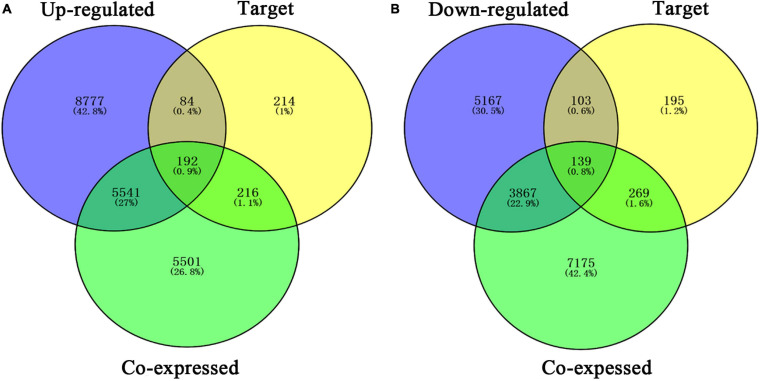
Esophageal squamous cell carcinoma (ESCC) is the major histological type of esophageal cancers worldwide. Transcription factor PTTG1 was seen highly expressed in a variety of tumors and was related to the degree of tumor differentiation, invasion, and metastasis. However, the clinical significance of PTTG1 had yet to be verified, and the mechanism of abnormal PTTG1 expression in ESCC was not clear. In this study, the comprehensive analysis and evaluation of PTTG1 expression in ESCC were completed by synthesizing in-house immunohistochemistry (IHC), clinical sample tissue RNA-seq (in-house RNA-seq), public high-throughput data, and literature data. We also explored the possible signaling pathways and target genes of PTTG1 in ESCC by combining the target genes of PTTG1 (displayed by ChIP-seq), differentially expressed genes (DEGs) of ESCC, and PTTG1-related genes, revealing the potential molecular mechanism of PTTG1 in ESCC. In the present study, PTTG1 protein and mRNA expression levels in ESCC tissues were all significantly higher than in non-cancerous tissues. The pool standard mean difference (SMD) of the overall PTTG1 expression was 1.17 (95% CI: 0.72-1.62, P < 0.01), and the area under curve (AUC) of the summary receiver operating characteristic (SROC) was 0.86 (95% CI: 0.83-0.89). By combining the target genes displayed by ChIP-seq of PTTG1, DEGs of ESCC, and PTTG1-related genes, it was observed that PTTG1 may interact with these genes through chemokines and cytokine signaling pathways. By constructing a protein-protein interaction (PPI) network and combining ChIP-seq data, we obtained four PTTG1 potential target genes, SPTAN1, SLC25A17, IKBKB, and ERH. The gene expression of PTTG1 had a strong positive correlation with SLC25A17 and ERH, which suggested that PTTG1 might positively regulate the expression of these two genes. In summary, the high expression of PTTG1 may play an important role in the formation of ESCC. These roles may be completed by PTTG1 regulating the downstream target genes SLC25A17 and ERH.

Keywords: PTTG1; RNA sequencing; esophageal squamous cell carcinoma; tissue microarray; transcription factor.


Conflict of interest statement

The authors declare that the research was conducted in the absence of any commercial or financial relationships that could be construed as a potential conflict of interest.

Figures

FIGURE 1
FIGURE 1
PTTG1 protein expression elevated in esophageal squamous cell carcinoma (ESCC) tissue chip assessed by in-house immunohistochemistry. (A,B) Immunohistochemical staining of PTTG1 protein in non-cancerous esophageal tissues from different cases. Both cases were scored as 0 (×400); (C–F) ESCC tissues (×400). The scores of case (C) and case (D) were both 6. The score was recorded as 8 for case (E) and 12 for case (F); (G) box diagram; (H) receiver operating characteristic (ROC) curve; AUC, area under the curve.
FIGURE 2
FIGURE 2
In-house RNA-seq confirmed PTTG1 overexpression in ESCC tissues. (A) Heatmap of differentially expressed genes (DEGs). (B) Scatter plot. (C) ROC curve.
FIGURE 3
FIGURE 3
Flow chart of data collection for this study.
FIGURE 4
FIGURE 4
Expression of PTTG1 mRNA upregulates in ESCC based on high-throughput RNA-seq of public database. GPL570 (A,G), GPL571 (B,H), GSE23400 (C,I), GSE32424 (D,J), GSE45168 (E,K), and GSE70409 (F,L). The cancer genome atlas (TCGA) dot plot and ROC curve (M–O) KM survival analysis curve based on TCGA data from KMplot database; the median expression value of PTTG1 was applied as cut-off value to classify patients into low and high expression groups.
FIGURE 5
FIGURE 5
Comprehensive analysis further confirmed that PTTG1 was significantly overexpressed in ESCC. (A) Summary receiver operating characteristic (SROC) curve. (B) Funnel chart of publication bias. AUC, area under curve.
FIGURE 6
FIGURE 6
Venn diagrams of overlapping PTTG1-related genes, target genes, and upregulated genes (A) and downregulated genes (B). Upregulated, upregulated differential genes; downregulated, downregulated differential genes; target, target genes of PTTG1 in ChIP-seq.
FIGURE 7
FIGURE 7
Gene ontology (GO) function annotation and Kyoto Encyclopedia of Genes and Genomes (KEGG) pathway analysis of upregulated overlapping genes. (A) Biological process. (B) Cell composition. (C) Molecular function. (D) KEGG pathway analysis.
FIGURE 8
FIGURE 8
GO function annotation and KEGG pathway analysis of downregulated overlapping genes. (A) Biological process. (B) Cell composition. (C) Molecular function. (D) KEGG pathway analysis.
FIGURE 9
FIGURE 9
Protein–protein interaction (PPI) network of overlapping genes. (A) PPI network of upregulated overlapping genes. (B) PPI network of downregulated overlapping genes.
FIGURE 10
FIGURE 10
Peaks of target genes binding to PTTG1 in ChIP-seq based on the Cistrome DB database. The peak signals of each target genes are colored red. (A) SPTAN1; (B) SLC25A17; (C) IKBKB; (D) ERH.
FIGURE 11
FIGURE 11
Pearson correlation analysis of PTTG1 and its target genes. (A) Pearson correlation between PTTG1 and SLC25A17. (B) Pearson correlation between PTTG1 and ERH.

Similar articles

Cited by

References

    1. Ackermann A., Schrecker C., Bon D., Friedrichs N., Bankov K., Wild P., et al. (2019). Downregulation of SPTAN1 is related to MLH1 deficiency and metastasis in colorectal cancer. PLoS One 14:e0213411. 10.1371/journal.pone.0213411 - DOI - PMC - PubMed
    1. Agrimi G., Russo A., Scarcia P., Palmieri F. (2012). The human gene SLC25A17 encodes a peroxisomal transporter of coenzyme A, FAD and NAD+. Biochem. J. 443 241–247. 10.1042/bj20111420 - DOI - PubMed
    1. Arenas M. A. R., Whitsett T. G., Aronova A., Henderson S. A., LoBello J., Habra M. A., et al. (2018). Protein Expression of PTTG1 as a Diagnostic Biomarker in Adrenocortical Carcinoma. Ann. Surg. Oncol. 25 801–807. 10.1245/s10434-017-6297-6291 - DOI - PubMed
    1. Arnal M. J. D., Arenas Áf, Arbeloa Ál. (2015). Esophageal cancer: Risk factors, screening and endoscopic treatment in Western and Eastern countries. World J. Gastroenterol. 21 7933–7943. 10.3748/wjg.v21.i26.7933 - DOI - PMC - PubMed
    1. Ersvær E., Kildal W., Vlatkovic L., Cyll K., Pradhan M., Kleppe A., et al. (2020). Prognostic value of mitotic checkpoint protein BUB3, cyclin B1, and pituitary tumor-transforming 1 expression in prostate cancer. Mod. Pathol. 33 905–915. 10.1038/s41379-019-0418-412 - DOI - PMC - PubMed