

Incidentally Detected Sternal Foramen during an Urgent Surgical Revascularization: A Case Report
- PMID: 33552200
- PMCID: PMC7825463
- DOI: 10.18502/jthc.v15i2.4189
Incidentally Detected Sternal Foramen during an Urgent Surgical Revascularization: A Case Report
Abstract
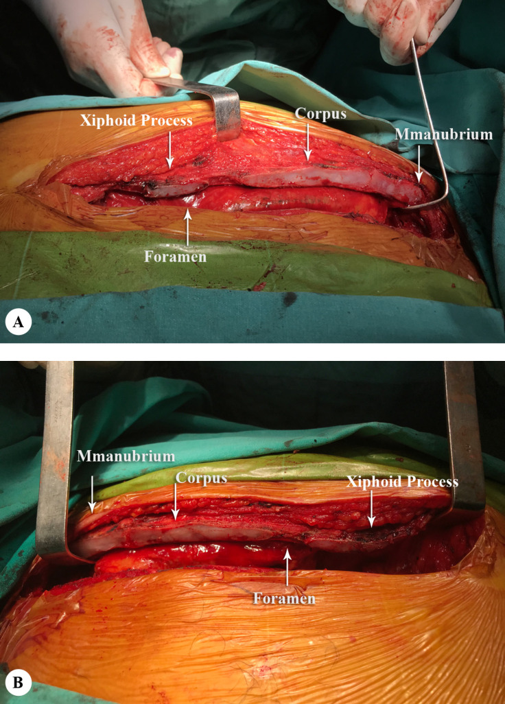
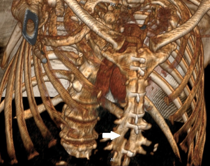
The sternal foramen is an anatomical variation at the lower third of the sternum which carries the risk of life-threatening complications such as pneumothorax. It is usually asymptomatic and can be misinterpreted as an acquired lesion. The sternum is close to the mediastinal structures; the sternal foramen, thus, leaves the lung, heart, and great vessels unprotected during invasive procedures. A 61-year-old male patient was admitted to our emergency department with sudden-onset chest pain. Acute coronary syndrome was diagnosed by the cardiology department. Coronary angiography confirmed the diagnosis of coronary artery disease, and the patient underwent urgent coronary artery bypass grafting. During the exploration, sternotomy was performed with a proper incision. Two sternal images were obtained via the median incision, and a sternal foramen was detected intraoperatively. To the best of our knowledge, this is the first case of sternotomy to undergo open-heart surgery for coronary heart disease and to be diagnosed with the sternal foramen intraoperatively. It is of vital importance that surgeons and interventionists recognize the sternal foramen, which leaves the mediastinal structures unprotected, and take early precautions.
Keywords: Anatomic variation; Coronary artery disease; Sternotomy.
Copyright © 2020 Tehran University of Medical Sciences.
Figures

References
-
- Babinski MA, de Lemos L, Babinski MS, Gonçalves MV, De Paula RC, Fernandes RM. Frequency of sternal foramen evaluated by MDCT: a minor variation of great relevance. Surg Radiol Anat. 2015;37:287–291. - PubMed
-
- Yekeler E, Tunaci M, Tunaci A, Dursun M, Acunas G. Frequency of sternal variations and anomalies evaluated by MDCT. AJR Am J Roentgenol. 2006;186:956–960. - PubMed
-
- Cooper PD, Stewart JH, McCormick WF. Development and morphology of the sternal foramen. Am J Forensic Med Pathol. 1988;9:342–347. - PubMed
-
- Moore MK, Stewart JH, McCormick WF. Anomalies of the human chest plate area. Radiographic findings in a large autopsy population. Am J Forensic Med Pathol. 1988;9:348–354. - PubMed
Publication types
LinkOut - more resources
Full Text Sources