

Primary undifferentiated pleomorphic cardiac sarcoma presenting as right heart failure
- PMID: 33552334
- PMCID: PMC7847826
- DOI: 10.1016/j.radcr.2021.01.016
Primary undifferentiated pleomorphic cardiac sarcoma presenting as right heart failure
Abstract
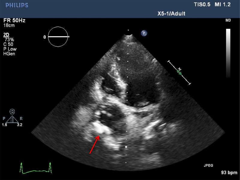
Right-sided heart failure is a common sequela of left heart failure and seldom presents as a primary disorder. The differential diagnosis of right heart failure includes a cardiac tumor. Cardiac malignancies are rare tumors with an overall poor prognosis. We evaluated a 69-year-old man who presented with a 3-week history of progressive lower extremity swelling, ascites, and scrotal swelling. Laboratory studies were significant only for mildly elevated liver function tests. CT scan of the abdomen and pelvis showed ascites, hepatic swelling, and a bland clot in the inferior vena cava extending from the level of the kidneys to the right atrium. A large mass originating from the right atrium was identified, and biopsy confirmed an undifferentiated pleomorphic cardiac sarcoma. Given the extensive tumor and clot burden, he was not an operative candidate. He developed portal hypertension with esophageal varices and expired due to variceal bleeding.
Keywords: Cardiac sarcoma; Right heart failure; Undifferentiated sarcoma.
© 2021 Published by Elsevier Inc. on behalf of University of Washington.
Figures


References
-
- Harjola V-P, Mebazaa A, Čelutkienė J. Contemporary management of acute right ventricular failure: a statement from the Heart Failure Association and the Working Group on Pulmonary Circulation and Right Ventricular Function of the European Society of Cardiology. Eur J Heart Fail. 2016;18:226–241. - PubMed
-
- Voelkel NF, Quaife RA, Leinwand LA. Right ventricular function and failure. Circulation. 2006;114:1883–1891. - PubMed
-
- Reardon MJ, DeFelice CA, Sheinbaum R. Cardiac autotransplant for surgical treatment of a malignant neoplasm. Ann Thorac Surg. 1999;67:1793–1795. - PubMed
-
- Lam K, Dickens P, Chan A. Tumors of the heart. A 20-year experience with a review of 12, 485 consecutive autopsies. Arch Pathol Lab Med. 1993;117:1027–1031. - PubMed
Publication types
LinkOut - more resources
Full Text Sources
Other Literature Sources
