. 2021 Jan 31;10(2):e1245.
doi: 10.1002/cti2.1245. eCollection 2021.

Pattern of circulating SARS-CoV-2-specific antibody-secreting and memory B-cell generation in patients with acute COVID-19

Affiliations

Pattern of circulating SARS-CoV-2-specific antibody-secreting and memory B-cell generation in patients with acute COVID-19

Maria Byazrova et al. Clin Transl Immunology. .

Abstract

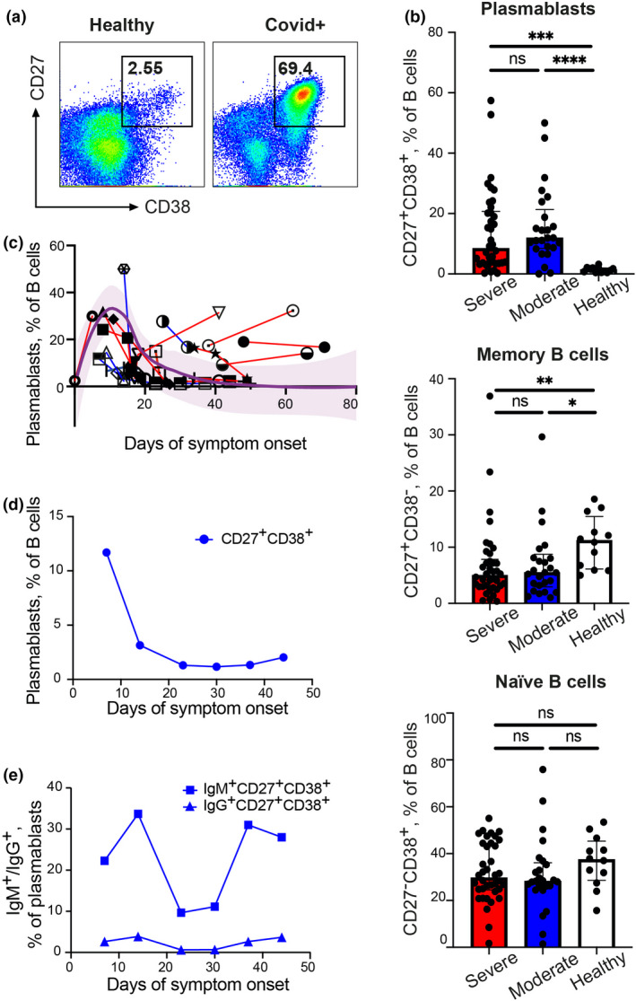
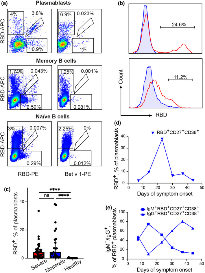
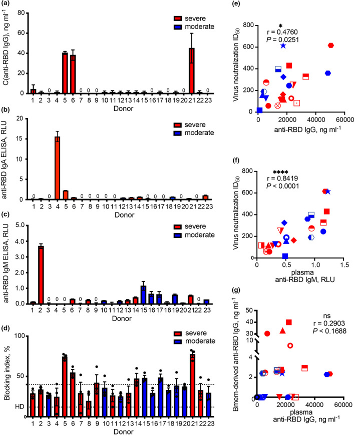
Objectives: To predict the spread of coronavirus disease (COVID-19), information regarding the immunological memory for disease-specific antigens is necessary. The possibility of reinfection, as well as the efficacy of vaccines for COVID-19 that are currently under development, will largely depend on the quality and longevity of immunological memory in patients. To elucidate the process of humoral immunity development, we analysed the generation of plasmablasts and virus receptor-binding domain (RBD)-specific memory B (Bmem) cells in patients during the acute phase of COVID-19.

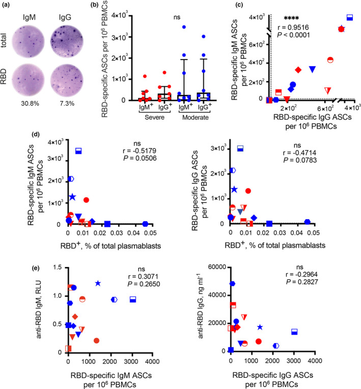
Methods: The frequencies of RBD-binding plasmablasts and RBD-specific antibody-secreting cells (ASCs) in the peripheral blood samples collected from patients with COVID-19 were measured using flow cytometry and the ELISpot assay.

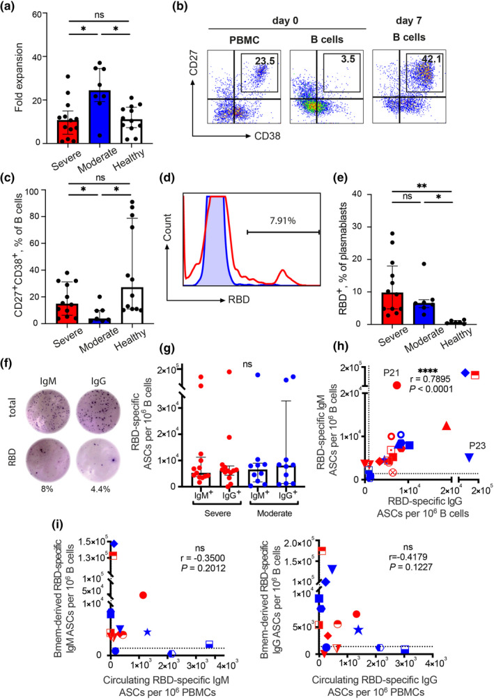
Results: The acute phase of COVID-19 was characterised by the transient appearance of total as well as RBD-binding plasmablasts. ELISpot analysis indicated that most patients exhibited a spontaneous secretion of RBD-specific ASCs in the circulation with good correlation between the IgG and IgM subsets. IL-21/CD40L stimulation of purified B cells induced the activation and proliferation of Bmem cells, which led to the generation of plasmablast phenotypic cells as well as RBD-specific ASCs. No correlation was observed between the frequency of Bmem cell-derived and spontaneous ASCs, suggesting that the two types of ASCs were weakly associated with each other.

Conclusion: Our findings reveal that SARS-CoV-2-specific Bmem cells are generated during the acute phase of COVID-19. These findings can serve as a basis for further studies on the longevity of SARS-CoV-2-specific B-cell memory.

Keywords: SARS‐CoV‐2; antibody‐secreting cell; memory B cell; plasmablast; virus‐neutralising antibodies.


Conflict of interest statement

The authors declare no conflict of interest.

Figures

Figure 1
Figure 1
Increased plasmablast frequency in patients with COVID‐19. (a) Flow cytometry dot plots showing plasmablast detection in a representative HD (left) and patient with severe COVID‐19 (right). Numbers inside the plots indicate the percentage of events specific to respective gates; 106 B‐cell events were acquired for each plot. (b) Plasmablast (left), memory B‐cell (middle) and naïve B‐cell (right) frequencies within the total CD19+ population in samples collected from HDs and patients with severe and moderate COVID‐19. (c) Dynamic changes in plasmablast frequencies in samples collected from 16 patients based on the number of days after symptom onset. Symbols connected by solid lines represent two to four time points considered for each patient. LOESS‐smoothed line and 95% confidence intervals are shown for 10 patients. (d, e) Longitudinal analysis of total (d) or IgM+/IgG+ (e) plasmablast response in a patient with moderate COVID‐19 (P24). Results are shown for individual samples (symbols) from HDs (n = 12), and moderate (n = 25) and severe (n = 38) COVID‐19 cases. Data are presented as median ± IQR. Asterisks indicate significant difference between groups determined using the Kruskal–Wallis test, *P < 0.05, **P < 0.01, ***P < 0.001, ****P < 0.0001, ns = not significant. HD, healthy donor; IQR, interquartile range.
Figure 2
Figure 2
Quantification of RBD‐specific B cells in fresh blood samples collected from patients with COVID‐19. (a) Representative flow cytometry dot plots showing double discrimination of RBD+ cells in plasmablast (top row), memory B‐cell (middle row) and naïve B‐cell (bottom row) subsets. Plasmablasts, memory B cells and naïve B cells were gated as CD27+CD38+, CD27+CD38 and CD27, respectively; 800 000 B‐cell events were acquired for each plot. (b) Representative bimodal (top panel) or unimodal (bottom panel) phycoerythrin‐labelled RBD staining histograms. For comparison, binding to the irrelevant protein Betv1‐PE is shown. (c) RBD+ frequencies within plasmablast population from HDs and patients with moderate (n = 22) and severe (n = 31) COVID‐19. (d, e) Dynamic changes in total (d) or IgM+/IgG+ (e) RBD‐specific plasmablast populations in sample obtained from P24 (patient with moderate COVID‐19). Results are shown for individual samples (symbols) from HDs (n = 12), and moderate (n = 25) and severe (n = 38) COVID‐19 cases. Data are presented as median ± IQR. Asterisks indicate significant difference between groups determined using the Kruskal–Wallis test, ****P < 0.0001, ns = not significant. HD, healthy donor; IQR, interquartile range; RBD, receptor‐binding domain.
Figure 3
Figure 3
ELISpot assay of circulating ASCs. (a) Representative ELISpot showing RBD‐specific circulating ASCs. Fresh PBMCs were incubated on ELISpot plates for 16 h to detect cells secreting total (top row) or RBD‐specific (bottom row) IgMs (left column) or IgGs (right column). The wells shown contained 104 PBMCs obtained from patients with COVID‐19. The percentages indicated beside the wells represent the frequencies of antigen‐specific ASCs relative to the total number of IgMs or IgGs. (b) RBD‐specific ASCs per 106 PBMCs collected from patients with severe (n = 7) and moderate (n = 8) COVID‐19. The dotted lines indicate the threshold for a positive RBD‐specific ASC response (20 spots per 106 PBMCs). (c) Scatter plot of IgM vs. IgG RBD‐specific ASCs. The dotted lines indicate the threshold for a positive RBD‐specific ASC response (20 spots per 106 PBMCs). (d) Frequencies of circulating RBD+ plasmablasts (percentage of total PBMCs) determined using flow cytometry, and RBD‐specific ASC IgMs (left panel) or IgGs (right panel) determined using the ELISpot assay. (e) Frequencies of circulating RBD‐specific ASC IgMs (left panel) or IgGs (right panel) determined using the ELISpot assay, and the levels of plasma RBD‐specific IgM (left panel) or IgG (right panel) determined using the ELISA. Data are presented as median ± IQR. ASC, antibody‐secreting cell; HD, healthy donor; PBMC, peripheral blood mononuclear cell; IQR, interquartile range; RBD, receptor‐binding domain.
Figure 4
Figure 4
CD40L/IL‐21‐induced cell expansion and activation and ASC generation in purified B cells. (a) Fold expansion after 7 days of IL‐21/CD40L stimulation of B cells from HDs (n = 12), and moderate (n = 10) and severe (n = 13) COVID‐19 cases. (b) Representative flow plots depicting the generation of stimulated B cells in vitro. Fresh PBMCs (left), immunomagnetically purified B cells (middle), and B cells after stimulation for 7 days (right) were analysed using flow cytometry. (c) In vitro generation of CD27+CD38+ B cells after 7 days of IL‐21/CD40L stimulation in HDs and patients with moderate and severe COVID‐19. (d) Representative flow plot of RBD+CD27+CD38+ B cells after IL‐21/CD40L stimulation for 7 days; 500 000 B‐cell events were acquired. (e) In vitro generation of RBD+CD27+CD38+ B cells after IL‐21/CD40L stimulation for 7 days. (f) Representative ELISpot showing RBD‐specific Bmem cell‐derived ASCs. Purified B cells were stimulated with IL‐21/CD40L for 7 days and then incubated on ELISpot plates for 16 h to detect cells secreting total (top row) or RBD‐specific (bottom row) IgMs (right column) or IgGs (left column). The percentages indicated beside the wells represent the frequencies of antigen‐specific ASCs relative to the total number of IgMs or IgGs. The wells shown contained 104 purified B cells obtained from patients with COVID‐19. (g) RBD‐specific Bmem cell‐derived ASCs per 106 PBMCs in patients with severe (n = 13) and moderate (n = 10) COVID‐19. (h) Scatter plot of RBD‐specific IgG vs. IgM ASCs after 7 days of IL‐21/CD40L stimulation. The dotted lines indicate the threshold for a positive RBD‐specific ASC response (220 for IgG spots and 1400 for IgM spots per 106 B cells). (i) Scatter plot of circulating vs. Bmem cell‐derived RBD‐specific IgM (left panel) or IgG (right panel) ASCs. Data are presented as median ± IQR. Asterisks indicate significant difference between groups determined using the Kruskal–Wallis test, *P < 0.05, **P < 0.01, ****P < 0.0001, ns = not significant. ASC, antibody‐secreting cell; Bmem cell, memory B cell; HD, healthy donor; RBD, receptor‐binding domain.
Figure 5
Figure 5
Activity of anti‐SARS‐CoV‐2 antibodies derived from cultures of CD40L/IL‐21‐stimulated B cells. (a–c) Production of RBD‐specific IgGs (a), IgAs (b) or IgMs (c) in cultures of IL‐21/CD40L‐stimulated B cells obtained from different patients with COVID‐19 evaluated using ELISA. The dotted line indicates the mean level of total IgGs observed in the HD group. The results of three separate experiments are presented for all patients. (d) Antibody‐mediated neutralisation assay of HIV‐1‐based virions pseudotyped with spike proteins of SARS‐CoV‐2. Supernatants from IL‐21/CD40L‐stimulated B‐cell cultures were used as a source of antibodies. The bottom dotted line indicates the mean level of virus neutralisation observed in the HD group. The upper dotted line indicates a cut‐off value for the virus neutralisation test. The results of three separate experiments are presented for all patients. (e‐f) Spearman’s correlation between virus neutralisation half‐maximal inhibitory plasma dilution (ID50) values and the levels of plasma anti‐RBD IgG (e) or IgM (f). (g) Scatter plot of Bmem cell‐derived vs. plasma anti‐RBD IgG. C, concentration; HD, healthy donor; ID, inhibitory dilution; OD, optical density; RBD, receptor‐binding domain

References

    1. Shah VK, Firmal P, Alam A, Ganguly D, Chattopadhyay S. Overview of immune response during SARS‐CoV‐2 infection: lessons from the past. Front Immunol 2020; 11: 1949. - PMC - PubMed
    1. Moderbacher CR, Ramirez SI, Dan JM et al Antigen‐specific adaptive immunity to SARS‐CoV‐2 in acute COVID‐19 and associations with age and disease severity. Cell 2020; 183: 996–1012. - PMC - PubMed
    1. Walls AC, Park Y‐J, Tortorici MA, Wall A, McGuire AT, Veesler D. Structure, Function, and Antigenicity of the SARS‐CoV‐2 Spike Glycoprotein. Cell 2020; 181: 281–292. - PMC - PubMed
    1. Ju B, Zhang QI, Ge J et al Human neutralizing antibodies elicited by SARS‐CoV‐2 infection. Nature 2020; 584: 115–119. - PubMed
    1. Robbiani DF, Gaebler C, Muecksch F et al Convergent antibody responses to SARS‐CoV‐2 in convalescent individuals. Nature 2020; 584: 437–442. - PMC - PubMed

LinkOut - more resources