Review
. 2021 Jan 20:8:624096.
doi: 10.3389/fbioe.2020.624096. eCollection 2020.

Extracellular Vesicles in Musculoskeletal Pathologies and Regeneration

Affiliations
Review

Extracellular Vesicles in Musculoskeletal Pathologies and Regeneration

Marietta Herrmann et al. Front Bioeng Biotechnol. .

Abstract

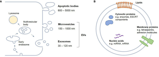
The incidence of musculoskeletal diseases is steadily increasing with aging of the population. In the past years, extracellular vesicles (EVs) have gained attention in musculoskeletal research. EVs have been associated with various musculoskeletal pathologies as well as suggested as treatment option. EVs play a pivotal role in communication between cells and their environment. Thereby, the EV cargo is highly dependent on their cellular origin. In this review, we summarize putative mechanisms by which EVs can contribute to musculoskeletal tissue homeostasis, regeneration and disease, in particular matrix remodeling and mineralization, pro-angiogenic effects and immunomodulatory activities. Mesenchymal stromal cells (MSCs) present the most frequently used cell source for EV generation for musculoskeletal applications, and herein we discuss how the MSC phenotype can influence the cargo and thus the regenerative potential of EVs. Induced pluripotent stem cell-derived mesenchymal progenitor cells (iMPs) may overcome current limitations of MSCs, and iMP-derived EVs are discussed as an alternative strategy. In the last part of the article, we focus on therapeutic applications of EVs and discuss both practical considerations for EV production and the current state of EV-based therapies.

Keywords: MSC; cell-free therapeutics; exosomes; extracellular vesicles; iMP; musculoskeletal diseases.


Conflict of interest statement

The authors declare that the research was conducted in the absence of any commercial or financial relationships that could be construed as a potential conflict of interest.

Figures

Figure 1
Figure 1
Biogenesis and content of extracellular vesicles (EVs). EVs are secreted by any cell type. (A) Based on the biogenesis route, different kinds of vesicles can be distinguished. Apoptotic bodies arise from membrane blebbing of apoptotic cells. Microvesicles derive from outward budding of the plasma membrane, while exosomes originate from the endo-lysosomal compartment and are released from the cell after fusion of multivesicular bodies with the plasma membrane. (B) EVs are enclosed by a lipid bilayer and carry membrane lipids and proteins, cytosolic proteins as well as nucleic acids, such as miRNAs and mRNAs. It is widely assumed that EV cargo mirrors the characteristics of the parent cell. ESCRT, endosomal sorting complexes required for transport.
Figure 2
Figure 2
iMP as an alternative cell source of EVs. Although MSCs can easily be maintained in cell culture enabling the production of large volumes of EV-conditioned medium, their proliferation and tri-lineage differentiation capacity declines during prolonged expansion and they are affected by donor variability. Induced pluripotent stem cells (iPSC) that derive from a somatic cell that underwent reprogramming with Myc-Oct3/4-Sox2-Klf4-expressing lentivirus represent an alternative cell source, with unlimited proliferation and differentiation potential that can overcome the donor variability issue. However, these benefits are surpassed by the technical issues related to iPSC maintenance and expansion in culture, as they require expensive media supplements, meticulous day-to-day inspections and handling, which make iPSC expansion in order to increase iPSC-EVs yield quite a challenging and expensive task. Alternatively, iPSC could be subjected to transdifferentiation into mesenchymal progenitor cells (iMP). These cells possess combined characteristics of iPSCs and MSCs: on one hand, they could be easily expanded and maintained in cell culture, like MSCs, and on the other hand, they inherit and retain the high proliferation and differentiation capacities of iPSCs.
Figure 3
Figure 3
Overview of different methodological approaches for isolation and purification of EVs. MSC-EVs secreted into a cell culture supernatant are collected as EV-conditioned medium (CM). All EV isolation protocols start with sequential low-speed centrifugation steps, to remove cell debris and large shed microvesicles. Next, the cleared CM can undergo either ultracentrifugation (1), ultrafiltration (2), precipitation with PEG (3), immune-immobilization (4), or size-exclusion chromatography (5) protocols resulting in isolated EV fractions. (1) CM is subjected to ultracentrifugation at 120 000 × g. For removing contaminating proteins, the EV pellet is additionally dissolved in PBS, and the ultracentrifugation step is repeated. (2) In case of ultrafiltration, the CM is passed through a membrane with pore sizes over 500 kDa MWCO by air pressure application. This results in concentrated CM with reduced serum protein and lipoprotein content. Next, the concentrated CM can either be subjected to a next round of ultrafiltration, using a membrane with pores ≤ 100 kDa MWCO, to retain and capture EVs, or, alternatively, it can undergo the ultracentrifugation procedure (1). For the PEG protocol (3), EVs are precipitated from the CM by 10% PEG in two rounds. The final EV pellet can be additionally purified with subsequent ultracentrifugation steps. For the immunocapture protocol (4), CD9-, CD63-, or CD81-conjugated magnetic beads are incubated with CM, to capture EVs, which are then immobilized by a magnet. After serial washing steps, the captured EVs are eluted from the beads. In case of size-exclusion chromatography (5), CM, or concentrated CM, is loaded on a resin-filled column, and the CM content (EVs and medium proteins) is fractionated according to their size. This approach allows isolation of uniformly-sized EV particles. All methods can be used as separate protocols or in various combinations, to fulfill different requirements for downstream analyses and applications utilizing EVs.
Figure 4
Figure 4
The role of EVs in musculoskeletal diseases—from pro-regenerative to detrimental effects. EVs are an important component of cell communication in musculoskeletal tissues. They can be produced by tissue resident cells as well as by cells, which invade in the process of disease progression and healing. As the parental cells, also EVs and their cargo are affected by microenvironmental changes. EVs can contribute to physiological and pathophysiological processes and thus critically influence tissue homeostasis and regeneration.

References

    1. Abbas M., Jesel L., Auger C., Amoura L., Messas N., Manin G., et al. . (2017). Endothelial microparticles from acute coronary syndrome patients induce premature coronary artery endothelial cell aging and thrombogenicity: role of the Ang II/AT1 receptor/NADPH oxidase-mediated activation of MAPKs and PI3-kinase pathways. Circulation 135, 280–296. 10.1161/CIRCULATIONAHA.116.017513 - DOI - PubMed
    1. Abramowicz A., Marczak L., Wojakowska A., Zapotoczny S., Whiteside T. L., Widlak P., et al. . (2018). Harmonization of exosome isolation from culture supernatants for optimized proteomics analysis. PLoS ONE 13:e0205496. 10.1371/journal.pone.0205496 - DOI - PMC - PubMed
    1. Adlerz K., Patel D., Rowley J., Ng K., Ahsan T. (2020). Strategies for scalable manufacturing and translation of MSC-derived extracellular vesicles. Stem Cell Res. 48:101978. 10.1016/j.scr.2020.101978 - DOI - PubMed
    1. Ahn S. Y., Park W. S., Kim Y. E., Sung D. K., Sung S. I., Ahn J. Y., et al. . (2018). Vascular endothelial growth factor mediates the therapeutic efficacy of mesenchymal stem cell-derived extracellular vesicles against neonatal hyperoxic lung injury. Exp. Mol. Med. 50:26. 10.1038/s12276-018-0055-8 - DOI - PMC - PubMed
    1. Alibhai F. J., Lim F., Yeganeh A., DiStefano P. V., Binesh-Marvasti T., Belfiore A., et al. . (2020). Cellular senescence contributes to age-dependent changes in circulating extracellular vesicle cargo and function. Aging Cell 19:e13103. 10.1111/acel.13103 - DOI - PMC - PubMed

LinkOut - more resources