. 2021 Jan 21:9:625805.
doi: 10.3389/fcell.2021.625805. eCollection 2021.

FKBP25 Regulates Meiotic Apparatus During Mouse Oocyte Maturation

Affiliations

FKBP25 Regulates Meiotic Apparatus During Mouse Oocyte Maturation

Danni Wang et al. Front Cell Dev Biol. .

Abstract

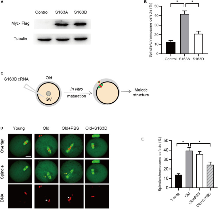
FK506 binding proteins 25 (FKBP25) has been shown to function in ribosome biogenesis, chromatin organization, and microtubule stability in mitosis. However, the role of FKBP25 in oocyte maturation has not been investigated. Here, we report that oocytes with FKBP25 depletion display abnormal spindle assembly and chromosomes alignment, with defective kinetochore-microtubule attachment. Consistent with this finding, aneuploidy incidence is also elevated in oocytes depleted of FKBP25. Importantly, FKBP25 protein level in old oocytes is significantly reduced, and ectopic expression of FKBP25 could partly rescue the aging-associated meiotic defects. In addition, by employing site-specific mutagenesis, we identify that serine 163 is a major, if not unique, phosphorylation site modulating the action of FKBP25 on meiotic maturation. In summary, our data indicate that FKBP25 is a pivotal factor for determining oocyte quality, and may mediate the effects of maternal aging on female reproduction.

Keywords: FKBP25; maternal aging; meiosis; oocyte; reproduction.


Conflict of interest statement

The authors declare that the research was conducted in the absence of any commercial or financial relationships that could be construed as a potential conflict of interest.

Figures

FIGURE 1
FIGURE 1
Cellular localization of FKBP25 in mouse oocyte. (A) Freshly collected oocytes of each stages were stained with FKBP25 antibody. Arrowheads point to the accumulation of FKBP25 signal. NC, negative control. (B) Metaphase oocytes were double labeled with FKBP25 antibody (red) and α-tubulin antibody (green), DNA was counterstained with Hoechst 33342 (blue). NC, negative control. Scale bar: 30 μm.
FIGURE 2
FIGURE 2
Effects of FKBP25 knockdown on oocyte maturation. (A) Western blot shows the knockdown efficiency of FKBP25 siRNA, 100 GV oocytes per sample. (B) Analysis the GVBD rate of control (n = 163 from 5 mice) and FKBP25-KD (n = 156 from 8 mice) oocytes. (C) Phase-contrast images of control and FKBP25 knockdown (FKBP25-KD) oocytes. Yellow arrowheads show the oocytes that fail to extrude polar bodies, pink arrowheads denote the oocytes with apparent symmetric division, blue asterisks point the oocytes arrested at GV stage, and white arrowheads indicate the oocytes with large polar bodies. Scale bar: 100 μm. (D) Magnified images for the abnormal oocytes shown in (C). (E) Analysis the Pb1 extrusion rate of control (n = 163 from 5 mice) and FKBP25-KD (n = 156 from 8 mice) oocytes. (F) The percentage of oocytes arrested at metaphase I stage after FKBP25-KD injection (n = 124 for control from 4 mice; n = 120 for FKBP25-KD from 6 mice). Data are expressed as mean ± SEM. **p < 0.01, ***p < 0.001 vs controls.
FIGURE 3
FIGURE 3
Morphological analysis of spindle and chromosome in FKBP25 knockdown oocytes. (A) Control and FKBP25 knockdown (FKBP25-KD) oocytes were stained with both α-tubulin antibody and PI. Disorganized spindles (yellow arrows) and misaligned chromosomes (white arrowheads) were frequently observed in FKBP25 knockdown (FKBP25-KD) oocytes. Scale bar: 30 μm. (B) Quantification of control (n = 124 from 4 mice) and FKBP25-KD (n = 120 from 6 mice) oocytes with spindle defects or chromosome misalignment. (C) Chromosome spread of control and FKBP25 knockdown (FKBP25-KD) oocytes. Chromosomes were labeled by Hoechst 33342 (blue) and kinetochores were stained with CREST (purple). Scale bar: 10 μm. (D) Quantification of aneuploidy in control (n = 51) and FKBP25 knockdown (n = 45) oocytes. Data are expressed as mean percentage ± SEM. **p < 0.01, ***p < 0.001 vs controls.
FIGURE 4
FIGURE 4
Depletion of FKBP25 in oocytes impairs kinetochore-microtubule attachments. (A) Metaphase oocytes in control and FKBP25-KD groups were double labeled with CREST and α-tubulin antibody. Representative confocal images are displayed. (B) Magnified images for the kinetochore-microtubule attachments in the oocytes shown in (A). (C) Quantitative analysis of K-MT attachments in oocytes (n = 52 for control; n = 56 for FKBP25-KD). (D) Control and FKBP25-KD oocytes were stained with anti-BubR1 antibody (green) and counterstained with PI to examine chromosomes (red). Scale bar: 10 μm. (E) BubR1 fluorescence intensity in control (n = 45) and FKBP25-KD (n = 50) oocytes were quantified. Data are expressed as mean percentage ± SEM. *p < 0.05 vs controls.
FIGURE 5
FIGURE 5
Expression of FKBP25 protein in oocytes from old mice. (A) Representative immunoblotting detecting the FKBP25 expression in oocytes from young and old mice (young, 100 oocytes from 3mice; old, 100 oocytes from 5 mice). (B) Confocal sections of young and old oocytes were immunolabeled with FKBP25 antibody (green) and counterstained with propidium iodide (PI) to visualize DNA (red). (C) Quantification analysis of FKBP25 fluorescence (27 young oocytes and 22 old oocytes for each experiment). Scale bar: 30 μm. *p < 0.05 vs controls.
FIGURE 6
FIGURE 6
FKBP25 overexpression alleviates maternal age-induced meiotic defects. (A) Schematic illustration of the FKBP25 overexpression experiments. (B,C) Immunoblotting analysis detecting the exogenous FKBP25 expression. (D) Oocytes were stained with α-tubulin and PI to detect spindle and chromosome morphology, respectively. Arrows indicate the disorganized spindles and arrowheads indicate the misaligned chromosomes. Scale bar: 30 μm. (E) Quantification of the spindle/chromosome defects in each group (young, n = 113 from 5 mice; old, n = 102 from 7 mice; old+PBS, n = 105 from 10 mice; old+FKBP25, n = 124 from 10 mice). (F) Chromosome spread of oocytes in each group. Chromosomes and kinetochores were stained with Hoechst 33342 (blue) and CREST (purple), respectively. Scale bar: 10 μm. (G) Quantification of aneuploidy in young (n = 39), old (n = 30), old+PBS (n = 32), and old+FKBP25 (n = 28) oocytes. Data are expressed as mean percentage ± SEM. *p < 0.05 vs controls.
FIGURE 7
FIGURE 7
FKBP25-S163D mutant partly rescues meiotic defects in old oocytes. (A) Immunoblotting analysis detecting the expression of two FKBP25 mutants. (B) The histogram shows the effects of FKBP phosphorylation mutants on meiotic apparatus in oocytes (n = 95 for each group). (C) Schematic diagram showing the experimental design process. (D) Oocytes in each group were labeled with α-tubulin antibody (green) for spindle and PI (red) for chromosome. Arrowheads point to the chromosome misalignment and arrows point to the spindle disorganization. Scale bar: 30 μm. (E) Quantification of oocytes with spindle/chromosome defects in each group (young, n = 121 from 5 mice; old, n = 108 from 7 mice; old+PBS, n = 102 from 10 mice; old+FKBP25, n = 132 from 10 mice). Data are expressed as mean percentage ± SEM. *p < 0.05 vs controls.

References

    1. Bonner M. K., Poole D. S., Xu T., Sarkeshik A., Yates J. R., III, Skop A. R. (2011). Mitotic spindle proteomics in Chinese hamster ovary cells. PLoS One 6:e20489. 10.1371/journal.pone.0020489 - DOI - PMC - PubMed
    1. Chambraud B., Belabes H., Fontaine-Lenoir V., Fellous A., Baulieu E. E. (2007). The immunophilin FKBP52 specifically binds to tubulin and prevents microtubule formation. FASEB J. 21 2787–2797. 10.1096/fj.06-7667com - DOI - PubMed
    1. Cheeseman I. M. (2014). The kinetochore. Cold Spring Harb. Perspect Biol. 6:a015826. 10.1101/cshperspect.a015826 - DOI - PMC - PubMed
    1. Chiang T., Schultz R. M., Lampson M. A. (2012). Meiotic origins of maternal age-related aneuploidy. Biol. Reprod. 86 1–7. 10.1095/biolreprod.111.094367 - DOI - PMC - PubMed
    1. Ciancimino L., Lagana A. S., Chiofalo B., Granese R., Grasso R., Triolo O. (2014). Would it be too late? A retrospective case-control analysis to evaluate maternal-fetal outcomes in advanced maternal age. Arch. Gynecol. Obstet. 290 1109–1114. 10.1007/s00404-014-3367-5 - DOI - PubMed

LinkOut - more resources