. 2021 Jan 21:9:626027.
doi: 10.3389/fcell.2021.626027. eCollection 2021.

Circular RNA CircCOL5A1 Sponges the MiR-7-5p/Epac1 Axis to Promote the Progression of Keloids Through Regulating PI3K/Akt Signaling Pathway

Affiliations

Circular RNA CircCOL5A1 Sponges the MiR-7-5p/Epac1 Axis to Promote the Progression of Keloids Through Regulating PI3K/Akt Signaling Pathway

Wenchang Lv et al. Front Cell Dev Biol. .

Abstract

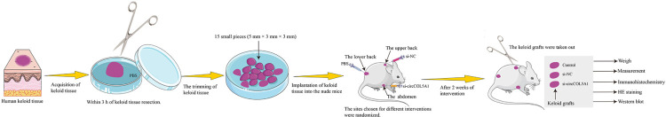
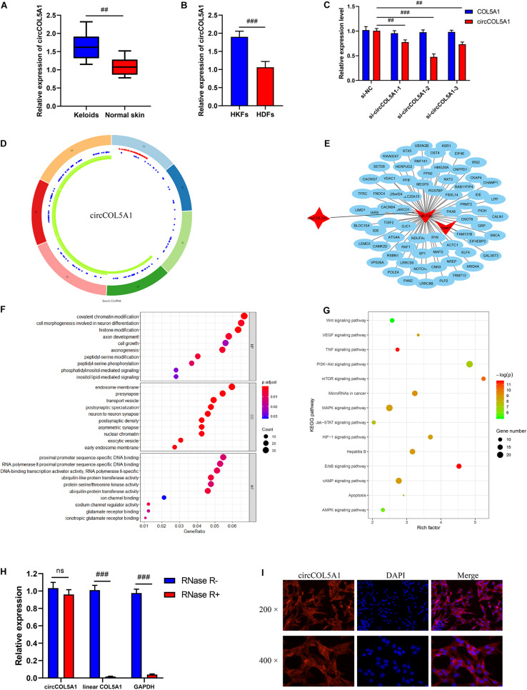
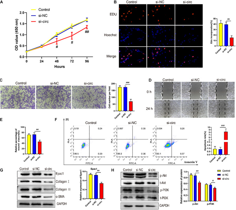
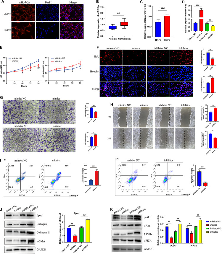
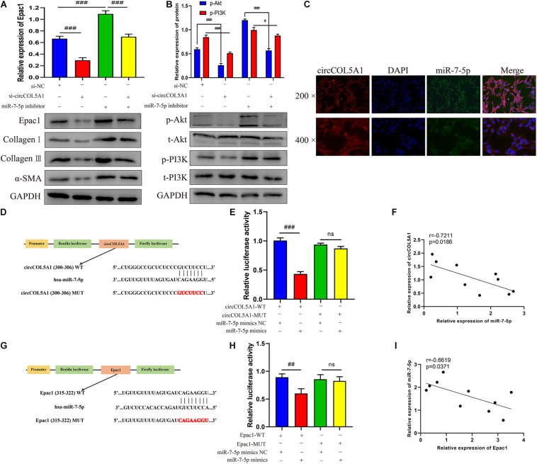
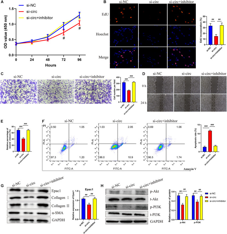
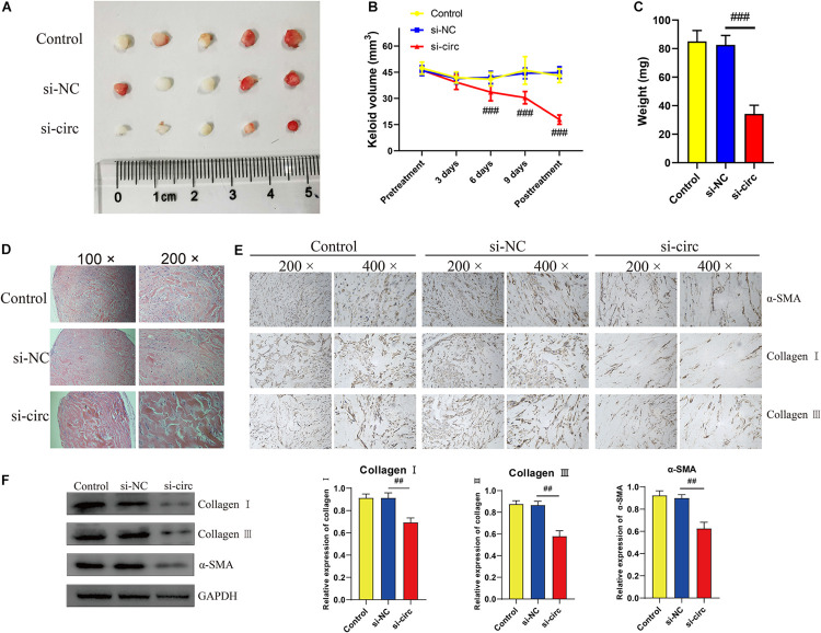
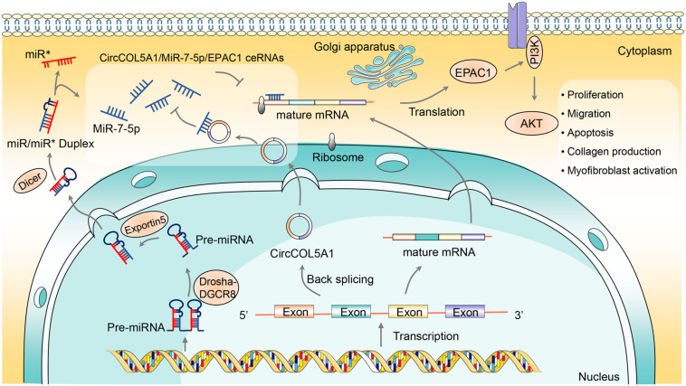
Keloids, as a result of abnormal wound healing in susceptible individuals, are characterized by the hyper-proliferation of fibroblasts and exaggerated deposition of extracellular matrix. Current surgical and therapeutic modalities provide limited satisfactory results. Growing evidence has highlighted the roles of circRNAs in acting as miRNA sponges. However, up to date, the regulatory mechanism of circRNAs in the pathological process of keloids has rarely been reported. In this study, cell proliferation, cell migration, flow cytometry, western blotting, fluorescence in situ hybridization, dual-luciferase activity, and immunohistochemistry assays were applied to explore the roles and mechanisms of the circCOL5A1/miR-7-5p/Epac1 axis in the keloid. The therapeutic potential of circCOL5A1 was investigated by establishing keloid implantation models. The RT-qPCR result revealed that circCOL5A1 expression was obviously higher in keloid tissues and keloid fibroblasts. Subsequent cellular experiments demonstrated that circCOL5A1 knockdown repressed the proliferation, migration, extracellular matrix (ECM) deposition, whereas promoted cell apoptosis, through the PI3K/Akt signaling pathway. Furthermore, RNA-fluorescence in situ hybridization (RNA-FISH) illustrated that both circCOL5A1 and miR-7-5p were located in the cytoplasm. The luciferase reporter gene assay confirmed that exact binding sites were present between circCOL5A1 and miR-7-5p, as well as between miR-7-5p and Epac1. Collectively, the present study revealed that circCOL5A1 functioned as competing endogenous RNA (ceRNA) by adsorbing miR-7-5p to release Epac1, which contributed to pathological hyperplasia of keloids through activating the PI3K/Akt signaling pathway. Our data indicated that circCOL5A1 might serve as a novel promising therapeutic target and represent a new avenue to understand underlying pathogenesis for keloids.

Keywords: Epac1; PI3K/Akt pathway; circCOL5A1; fibroblast; keloid; miR-7-5p.


Conflict of interest statement

The authors declare that the research was conducted in the absence of any commercial or financial relationships that could be construed as a potential conflict of interest.

Figures

SCHEME 1
SCHEME 1
Schematic illustration of implanting keloid tissues into nude mice to make the modeling process more intuitive and easier to understand.
FIGURE 1
FIGURE 1
Bioinformatics analysis and characterization of circCOL5A1. (A) The relative RNA levels of circCOL5A1 were evaluated by qRT-PCR between keloid tissues and normal skin. (B) The relative RNA levels of circCOL5A1 were evaluated by qRT-PCR between HKFs and HDFs. (C) The silent efficiency of circCOL5A1 was evaluated by qRT-PCR in HKFs transfected with si-NC or siRNAs, respectively. (D) The structure and binding sites of circCOL5A1. The red sites represented the microRNA response element. The blue sites represented RNA binding protein. The green sites represented an open reading frame. (E) Construction of circRNA-miRNA-mRNA network. GO (F) and KEGG (G) analysis of circCOL5A1 target genes. (H) The relative abundance of circCOL5A1 or linear COL5A1 in HKFs were detected by qRT-PCR after treatment with or without RNase R. (I) FISH assays were performed to observe the cellular location of circCOL5A1 (red) in HKFs (magnification, 200× and magnification, 400×). ##p < 0.01 and ###p < 0.001.
FIGURE 2
FIGURE 2
circCOL5A1 regulated HKFs proliferation, migration, apoptosis, and ECM deposition in vitro. (A) CCK-8 assays and (B) EdU assays were performed to assess the proliferation ability in HKFs transfected with the si-NC or si-circ, respectively. Magnification, 200×. (C) Transwell migration assays (magnification, 200×) and (D,E) wound healing assays (magnification, 20×) were applied for assessing the migration ability of HKFs transfected with the si-NC or si-circ, respectively. (F) Cell apoptosis was examined using flow cytometry. (G,H) The protein levels of collagen I, collagen III, α-SMA, and the protein phosphorylation levels of Akt and PI3K in HKFs transfected with si-NC or si-circ by western blot assays. Data was shown as mean ± SD. ns indicated no significance, #P < 0.05, ##P < 0.01, ###P < 0.001, vs. si-NC.
FIGURE 3
FIGURE 3
MiR-7-5p regulated HKFs proliferation, migration, apoptosis, and ECM deposition in vitro by targeting Epac1. (A) FISH assays were performed to observe the cellular location of miR-7-5p (red) in HKFs (magnification, 200× and magnification, 400×). (B) The relative RNA levels of miR-7-5p were evaluated by qRT-PCR between keloid tissues and normal skin. (C) The relative RNA levels of miR-7-5p were evaluated by qRT-PCR between HKFs and HDFs. (D) The transfection efficiency of miR-7-5p was evaluated by qRT-PCR in HKFs transfected with the miR-7-5p mimics or inhibitor, respectively. (E) CCK-8 assays and (F) EdU assays were performed to evaluate the proliferation ability in HKFs transfected with the miR-7-5p mimics or inhibitor, respectively. Magnification, 200×. (G) Transwell migration assays (magnification, 200×) and (H) wound healing assays (magnification, 20×) were applied for assessing the migration ability of HKFs transfected with the miR-7-5p mimics or inhibitor, respectively. (I) Cell apoptosis was examined using flow cytometry. (J,K) The protein levels of Epac1, collagen I, collagen III, α-SMA, and the protein phosphorylation levels of Akt and PI3K in HKFs transfected with miR-7-5p mimics or inhibitor by western blot assays. Data was shown as mean ± SD. ns indicated no significance, #P < 0.05, ##P < 0.01, ###P < 0.001, vs. NC.
FIGURE 4
FIGURE 4
circCOL5A1 served as a miRNA sponge of miR-7-5p to regulate Epac1 expression. (A,B) The protein levels of collagen I, collagen III, α-SMA, and the protein phosphorylation levels of Akt and PI3K in HKFs transfected with si-NC, si-circ, or si-circ + inhibitor were determined using western blot, respectively. (C) FISH assays were performed to observe the cellular location of circCOL5A1 (red) and miR-7-5p (green) in HKFs (magnification, 200× and magnification, 400×). (D) Schematic diagram of circCOL5A1-WT and circCOL5A1-MUT luciferase reporter vectors. (E) The relative luciferase activities were evaluated in HKFs after co-transfection with circCOL5A1-WT or circCOL5A1-MUT and mimics or NC, respectively. (F) Pearson correlation analysis was performed to evaluate the correlation between circCOL5A1 and miR-7-5p in keloid tissues. (G) Schematic diagram of miR-7-5p-WT and miR-7-5p-MUT luciferase reporter vectors. (H) The luciferase activity of reporter that carried WT rather than Mut 3′-UTR of Epac1 was markedly suppressed by miR-7-5p mimics. (I) Pearson correlation analysis was performed to evaluate the correlation between miR-7-5p and Epac1 in keloid tissues. Data was shown as mean ± SD. ns indicated no significance, #P < 0.05, ##P < 0.01, ###P < 0.001.
FIGURE 5
FIGURE 5
circCOL5A1 regulated HKFs proliferation, migration, apoptosis, and ECM deposition through circCOL5A1/miR-7-5p/Epac1 axis. (A) CCK-8 assays and (B) EdU assays were performed to evaluate the proliferation ability in HKFs transfected with the si-NC, si-circ, or si-circ + inhibitor, respectively. Magnification, 200×. (C) Transwell migration assays (magnification, 200×) and (D,E) wound healing assays (magnification, 20×) were applied for assessing the migration ability of HKFs transfected with the si-NC, si-circ, or si-circ + inhibitor, respectively. (F) Cell apoptosis was examined using flow cytometry. (G,H) The protein levels of collagen I, collagen III, α-SMA, and the protein phosphorylation levels of Akt and PI3K in HKFs transfected with si-NC, si-circ, or si-circ + inhibitor by western blot assays. Data was shown as mean ± SD. ns indicated no significance, #P < 0.05, ##P < 0.01, ###P < 0.001, vs. si-NC.
FIGURE 6
FIGURE 6
Downregulation of circCOL5A1 suppressed the growth of keloids and ECM deposition in vivo. (A) Images of subcutaneous keloid grafts in circCOL5A1 low expression group and control group. (B) The relative volume of keloid grafts was analyzed. (C) The weight of the keloid grafts was evaluated. (D) Representative images of HE staining of keloid nodules in different intervention groups (magnification, 100× and magnification, 200×). (E) The relative expression level of collagen I, collagen III, and α-SMA was observed in keloid grafts by IHC (magnification, 200× and magnification, 400×). (F) The protein levels of collagen I, collagen III, and α-SMA were evaluated in subcutaneous keloid grafts by western blot analysis. Data was shown as mean ± SD. ns indicated no significance, ##P < 0.01, ###P < 0.001, vs. si-NC.
FIGURE 7
FIGURE 7
The schematic diagram illustrates the role of circCOL5A1 as a ceRNA for miR-7-5p to release Epac1 in regulating the pathological phenotype of keloids. circCOL5A1 was generated by the head-to-tail splicing of COL5A1 in the nucleus and then functioned as ceRNA in the cytoplasm. circCOL5A1 reversed miR-7-5p-induced enhancement of HKFs biological function via the PI3K/Akt signaling pathway, including proliferation, migration, apoptosis, collagen production, and myofibroblast activation. Mechanismly, circRNAs could restore the expression of miR-7-5p target Epac1, forming the circCOL5A1/miR-7-5p/Epac1 regulating axis. The miR represented a segment of RNA on the pre-miRNA, and its position happened to be opposite that of the mature miRNA.

References

    1. Arjunan S., Gan S. U., Choolani M., Raj V., Lim J., Biswas A., et al. (2020). Inhibition of growth of Asian keloid cells with human umbilical cord Wharton’s jelly stem cell-conditioned medium. Stem Cell. Res. Ther. 11:78. 10.1186/s13287-020-01609-7 - DOI - PMC - PubMed
    1. Artemaki P. I., Scorilas A., Kontos C. K. (2020). Circular RNAs: a new piece in the colorectal cancer puzzle. Cancers 12:2464. 10.3390/cancers12092464 - DOI - PMC - PubMed
    1. Betarbet U., Blalock T. W. (2020). Keloids: a review of etiology, prevention, and treatment. J. Clin. Aesthet. Dermatol. 13 33–43. - PMC - PubMed
    1. Bieg D., Sypniewski D., Nowak E., Bednarek I. (2019). MiR-424-3p suppresses galectin-3 expression and sensitizes ovarian cancer cells to cisplatin. Arch. Gynecol. Obstet. 299 1077–1087. 10.1007/s00404-018-4999-7 - DOI - PMC - PubMed
    1. Christensen A. E., Selheim F., de Rooij J., Dremier S., Schwede F., Dao K. K., et al. (2003). cAMP analog mapping of Epac1 and cAMP kinase. Discriminating analogs demonstrate that Epac and cAMP kinase act synergistically to promote PC-12 cell neurite extension. J. Biol. Chem. 278 35394–35402. 10.1074/jbc.M302179200 - DOI - PubMed

LinkOut - more resources