. 2021 Jan 21:7:619859.
doi: 10.3389/fsurg.2020.619859. eCollection 2020.

Determination of the Optimum Excitation Wavelength for the Parathyroid Gland Using a Near-Infrared Camera

Affiliations

Determination of the Optimum Excitation Wavelength for the Parathyroid Gland Using a Near-Infrared Camera

Isao Tabei et al. Front Surg. .

Abstract

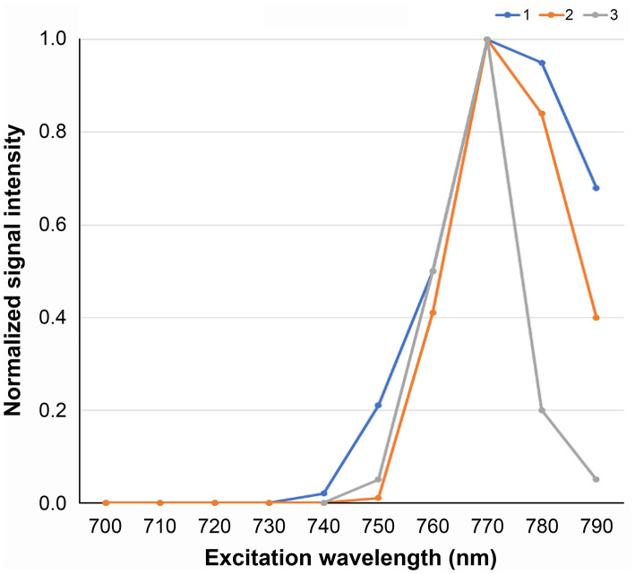
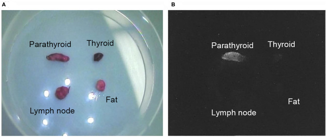
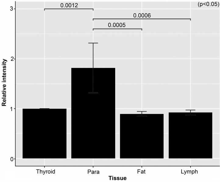
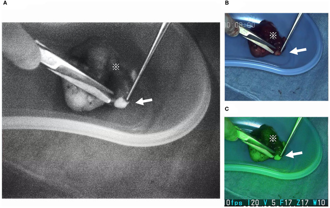
When performing thyroid/parathyroid surgery, difficulty detecting the parathyroid gland is a common experience because it is frequently mistaken with surrounding structures, including the thyroid gland, lymph nodes, and fat. To obtain successful surgical results, the auto fluorescent property of the parathyroid gland occurring at 820-830 nm has been used. Intraoperative visualization and detection by fluorescence enable protection of the gland from damage and unintended removal. Use of a near-infrared (NIR) camera has been proposed to indicate the parathyroid gland, but the devices and success rates have varied. This study aimed to define optimum excitation wavelength (EWL) by measuring the EWL of the parathyroid gland for its autofluorescence. Glands were exposed to EWL at 10-nm intervals from 670-790 nm with a light-emitting diode monochromator; autofluorescence intensity was recorded with a conventional NIR video camera. Autofluorescence intensity curves of three normal parathyroid glands were depicted; the optimum EWL was measured as 760-770 nm. Also, the illumination of the surrounding structures were compared at the optimum EWL. The auto fluorescent intensity of the parathyroid gland was 2-fold greater than for surrounding structures. This difference in fluorescence intensity should enable distinction of the parathyroid gland from surrounding structures. The clarification of the optimum EWL can guide refinements of the NIR camera for better surgical outcomes by improving detection of the parathyroid glands. Also, an understanding of optimum EWL should lead to developments for microscopic devices to unravel the still unknown mechanisms of the intrinsic autofluorescence of the parathyroid gland.

Keywords: ICG; autofluorescence; excitation wavelength; near-infrared camera; parathyroid gland.


Conflict of interest statement

Equipment used in this study were provided by Shimadzu Corporation and Hamamatsu Photonics K.K. The authors declare that the research was conducted in the absence of any commercial or financial relationships that could be construed as a potential conflict of interest.

Figures

Figure 1
Figure 1
Device used to measure the EWL characteristics of the parathyroid gland: (A) the fluorescence illumination was recorded with the LIGHTVISION NIR camera; (A,B) NIR EWL was projected to the extracted parathyroid gland from the monochromator SPG-120IR; (C) illumination data from the video recorded image were analyzed using ImageJ; the image is lightened by penlight to show the sample and projected EWL nm. EWL, excitation wavelength; NIR, near infrared.
Figure 2
Figure 2
Autofluorescence intensity characteristics for the three parathyroid glands. The optimum excitation wavelength for the parathyroid gland to autofluorescence absorbance wavelength of 800–850 nm was estimated to be 770 nm.
Figure 3
Figure 3
The pde-neoII NIR fluorescence imager C10935-400 used to monitor extracted samples. This device projects 760-nm NIR EWL by LED and collects the fluorescence illumination at 830 nm. This device is commercially sold to detect the sentinel lymph node in the breast cancer surgery with fluorescent light using indocyanine green. EWL, excitation wavelength; LED, light-emitting diode; NIR, near infrared.
Figure 4
Figure 4
Video image sample clip of the collected material. One sample set image clip of the parathyroid gland, thyroid, lymph node, and fat tissue are shown in one view to compare the relative illumination intensity ratio: (A) the optic visual light view under white light; (B) and fluorescence view.
Figure 5
Figure 5
Graph of the relative intensity of each tissue. The average relative intensity for the nine sample sets of the parathyroid (Para), fat, and lymph node (Lymph) compared with the thyroid is shown. As shown parathyroid was almost 2-fold brighter.
Figure 6
Figure 6
Extraction of the parathyroid gland at the back table in the operating room. The parathyroid gland (arrow) is often embedded with in the lower pole of the excised thyroid (※). The parathyroid gland is easily detected and located with the NIR camera. The projection source of the pde-neoII was used. The image is visualized using the LIGHTVISION NIR camera. This device enables three screens visualizing: (A) the NIR fluorescence, (B) the optic white light, and (C) the optic and modified NIR fluorescence screen visions projected simultaneously. NIR, near infrared.

References

    1. Bliss RD, Gauger PG, Delbridge LW. Surgeon's approach to the thyroid gland: surgical anatomy and the importance of technique. World J Surg. (2000) 24:891–97. 10.1007/s002680010173 - DOI - PubMed
    1. Marissa M, Natalie C, Gerald O, Stacey M, Michelle S, Erin R, et al. . Factors contributing to unintentional parathyroidectomy during thyroid surgery. Baylor Univ Med Cent Proc. (2020) 33:19–23. 10.1080/08998280.2019.1680911 - DOI - PMC - PubMed
    1. Kitai T, Inomoto T, Miwa M, Shikayama T. Fluorescence navigation with indocyanine green for detecting sentinel lymph nodes in breast cancer. Breast Cancer. (2005) 12:211–5. 10.2325/jbcs.12.211 - DOI - PubMed
    1. Watanabe J, Ishibe A, Suwa Y, Suwa H, Ota M, Kunisaki C, et al. . Indocyanine green fluorescence imaging to reduce the risk of anastomotic leakage in laparoscopic low anterior resection for rectal cancer: a propensity score-matched cohort study. Surg Endosc. (2020) 34:202–8. 10.1007/s00464-019-06751-9 - DOI - PubMed
    1. Alander JT, Kaartinen I, Laakso A, Pätilä T, Spillmann T, Tuchin VV, et al. . A review of indocyanine green fluorescent imaging in surgery. Int J Biomed Imaging. (2012) 2012:26. 10.1155/2012/940585 - DOI - PMC - PubMed