

Characteristics of Osteochondral Fractures Caused by Patellar Dislocation
- PMID: 33553448
- PMCID: PMC7841865
- DOI: 10.1177/2325967120974649
Characteristics of Osteochondral Fractures Caused by Patellar Dislocation
Abstract
Background: Literature describing the anatomic characteristics of osteochondral fractures (OCFs) in the knee joint after patellar dislocation is scarce.
Purpose: To describe the patterns of OCFs in the knee joint after acute or recurrent patellar dislocation in a sample of patients from 2 orthopaedic trauma centers.
Study design: Case series; Level of evidence, 4.
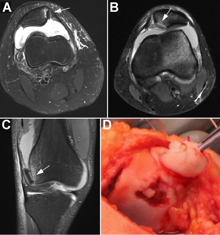
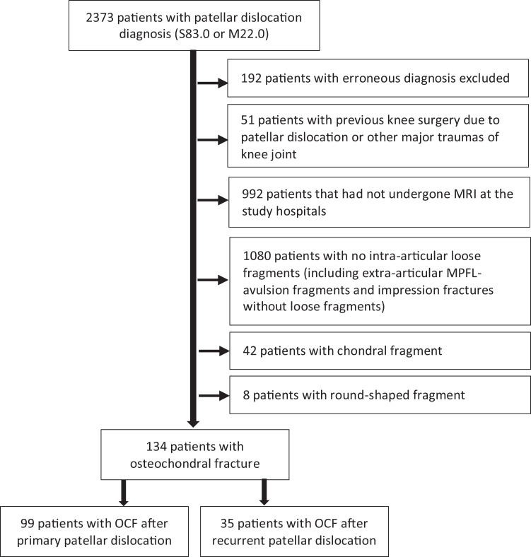
Methods: In this multicenter study, all patients who had International Classification of Diseases, 10th Revision, diagnostic codes S83.0 and M22.0 between 2012 and 2018 were screened. Of the 2181 patients with clinically diagnosed patellar dislocation, 1189 had undergone magnetic resonance imaging (MRI). Patients with diagnosed patellar dislocation and osteochondral fragment verified on MRI scans were included. Demographic and clinical data were collected from electronic patient records. OCF location and size were assessed from MRI scans. Results were further compared in subgroups by sex, skeletal maturity, and primary versus recurrent patellar dislocation.
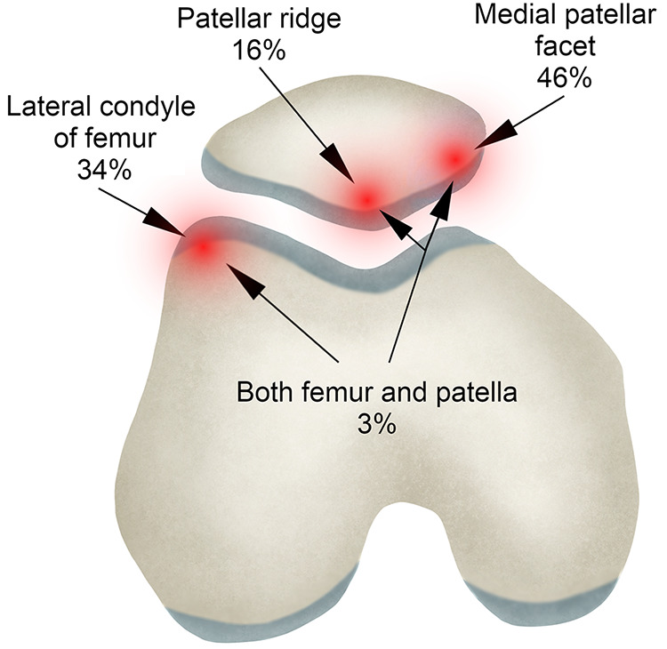
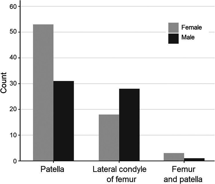
Results: An OCF was detected in 134 patients with injured knees, all of whom were included in the final analysis. It occurred in the patella in 63% of patients, in the lateral femoral condyle in 34%, and in both locations in 3%. The median OCF size was 146 mm2 (interquartile range, 105-262 mm2). There was no statistically significant difference in OCF size between patellar and lateral femoral condyle fractures. Patellar OCFs were more frequent in female than male patients (P = .009) and were larger after primary than recurrent dislocation (P = .040).
Conclusion: OCFs were mainly located in the medial facet of the patella and in the lateral femoral condyle, with these locations accounting for approximately two-thirds and one-third of all OCFs, respectively. Proportion of patellar OCF was higher in female than in male. Patellar OCFs may be larger after primary than recurrent dislocation.
Keywords: knee trauma; magnetic resonance imaging; osteochondral fracture; patellar dislocation.
© The Author(s) 2021.
Conflict of interest statement
The authors declared that there are no conflicts of interest in the authorship and publication of this contribution. AOSSM checks author disclosures against the Open Payments Database (OPD). AOSSM has not conducted an independent investigation on the OPD and disclaims any liability or responsibility relating thereto.
Figures
References
-
- Beasley LS, Vidal AF. Traumatic patellar dislocation in children and adolescents: treatment update and literature review. Curr Opin Pediatr. 2004;16:29–36. - PubMed
-
- Dejour D, Le Coultre B. Osteotomies in patello-femoral instabilities. Sports Med Arthrosc Rev. 2018;26:8–15. - PubMed
-
- Diederichs G, Issever AS, Scheffler S. MR imaging of patellar instability: injury patterns and assessment of risk factors. Radiographics. 2010;30:961–981. - PubMed
-
- Fithian DC, Paxton EW, Stone ML, et al. Epidemiology and natural history of acute patellar dislocation. Am J Sports Med. 2004;32:1114–1121. - PubMed
LinkOut - more resources
Full Text Sources
Other Literature Sources
