

A New Clinical Sign for Diagnosing Medial Meniscus Posterior Root Tear
- PMID: 33553453
- PMCID: PMC7841682
- DOI: 10.1177/2325967120975511
A New Clinical Sign for Diagnosing Medial Meniscus Posterior Root Tear
Abstract
Background: In the presence of medial meniscus posterior root tear (MMPRT), there is a possibility of reduced compression of meniscal tissue in hyperflexion as the intra-articular mobility of the meniscus increases. This phenomenon can be mimicked during clinical examination.
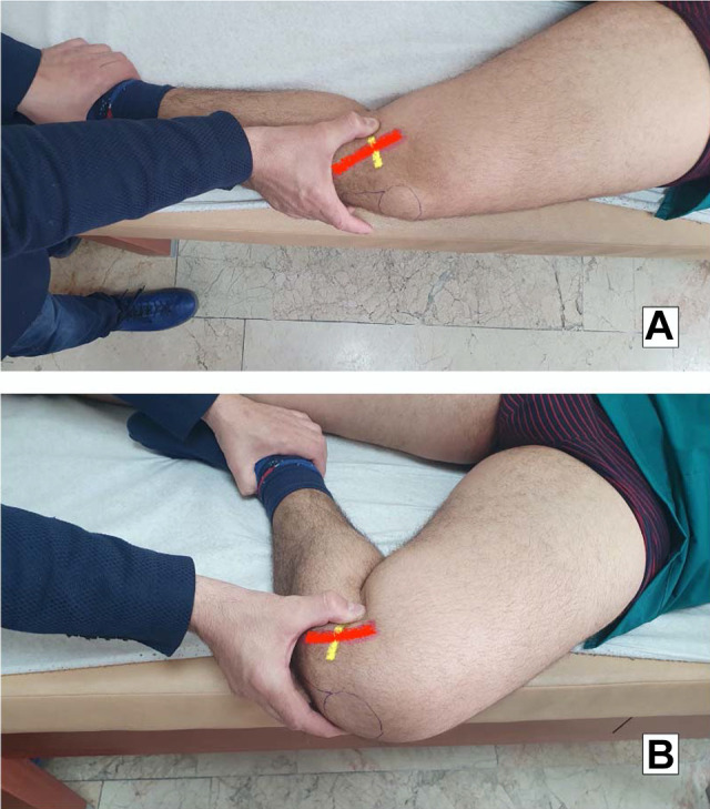
Purpose: To describe, evaluate, and validate the diagnostic performance of a new clinical indicator, the Akmese sign, for the diagnosis of an MMPRT.
Study design: Cohort study (diagnosis); Level of evidence, 2.
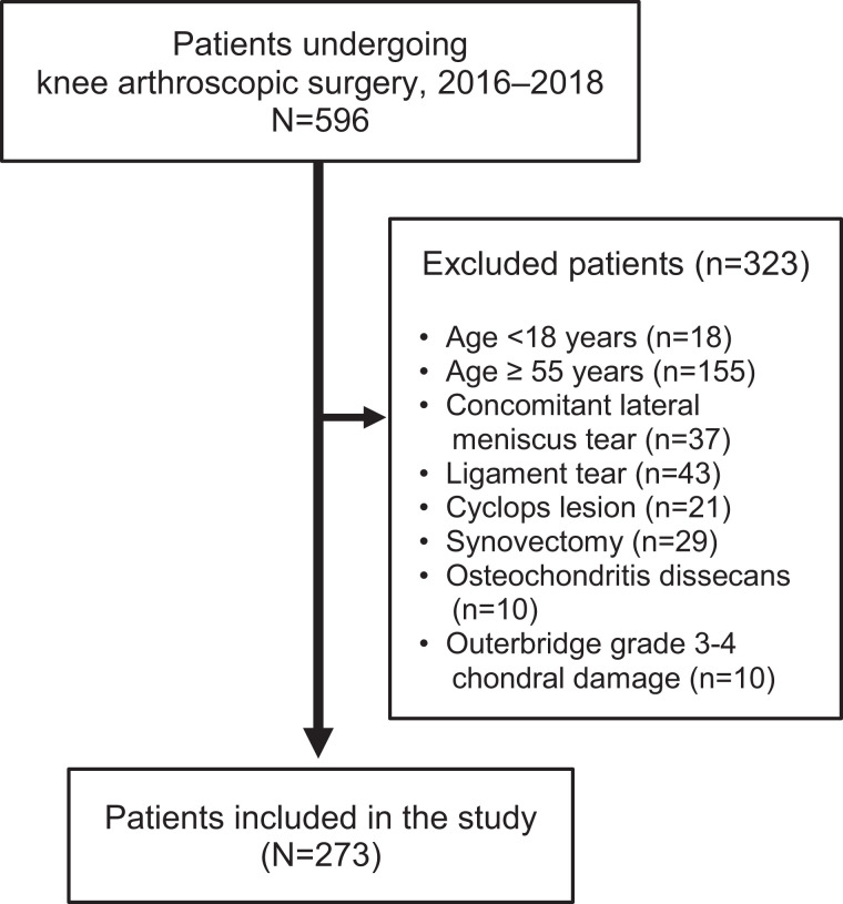
Methods: In this study, we prospectively enrolled patients aged 18 to 55 years who were scheduled for arthroscopic surgery after a diagnosis of medial meniscal lesion at a single institution between January 2016 and January 2018. All of the patients underwent preoperative examination for the Akmese sign. All surgeries were performed by a single surgeon with more than 5 years of experience in sports injury surgery, who was blinded to the Akmese sign results.
Results: A total of 273 patients with a mean age of 42.4 ± 5.3 years met the study criteria. The Akmese sign was identified as positive in 33 patients, and MMPRT was confirmed during arthroscopy in 36 cases. The performance parameters of the Akmese sign were a sensitivity of 86.1%, specificity of 99.1%, Youden index of 0.85, and kappa index of 0.88.
Conclusion: This study showed that the Akmese sign is a useful new physical examination test that can help clinicians distinguish MMPRTs from other meniscal medial meniscal pathology.
Keywords: Akmese sign; arthroscopy; knee; medial menisci; root tear; transtibial technique.
© The Author(s) 2021.
Conflict of interest statement
The authors declared that there are no conflicts of interest in the authorship and publication of this contribution. AOSSM checks author disclosures against the Open Payments Database (OPD). AOSSM has not conducted an independent investigation on the OPD and disclaims any liability or responsibility relating thereto.
Figures
References
-
- Ahn JH, Lee YS, Chang J-Y, Chang MJ, Eun SS, Kim SM. Arthroscopic all inside repair of the lateral meniscus root tear. Knee. 2009;16(1):77–80. - PubMed
-
- Allaire R, Muriuki M, Gilbertson L, Harner CD. Biomechanical consequences of a tear of the posterior root of the medial meniscus: similar to total meniscectomy. J Bone Joint Surg Am. 2008;90(9):1922–1931. - PubMed
-
- Bhatia S, LaPrade CM, Ellman MB, LaPrade RF. Meniscal root tears: significance, diagnosis, and treatment. Am J Sports Med. 2014;42(12):3016–3030. - PubMed
-
- Bin S-I, Kim J-M, Shin S-J. Radial tears of the posterior horn of the medial meniscus. Arthroscopy. 2004;20(4):373–378. - PubMed
-
- Choi C-J, Choi Y-J, Lee J-J, Choi C-H. Magnetic resonance imaging evidence of meniscal extrusion in medial meniscus posterior root tear. Arthroscopy. 2010;26(12):1602–1606. - PubMed
LinkOut - more resources
Full Text Sources
Other Literature Sources
Miscellaneous
