. 2021 Jan 22;6(8):2231-2249.
doi: 10.1016/j.bioactmat.2021.01.008. eCollection 2021 Aug.

Transplantation of engineered exosomes derived from bone marrow mesenchymal stromal cells ameliorate diabetic peripheral neuropathy under electrical stimulation

Affiliations

Transplantation of engineered exosomes derived from bone marrow mesenchymal stromal cells ameliorate diabetic peripheral neuropathy under electrical stimulation

Anamika Singh et al. Bioact Mater. .

Abstract

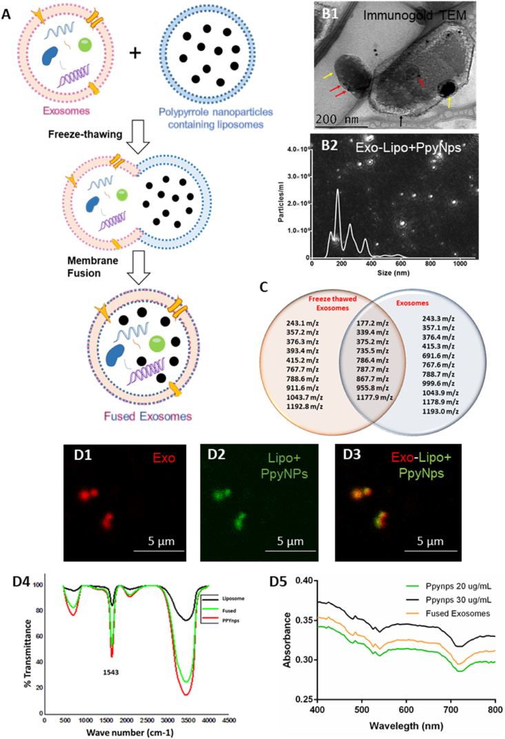
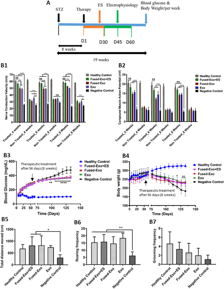
Diabetic peripheral neuropathy (DPN) is a long-term complication associated with nerve dysfunction and uncontrolled hyperglycemia. In spite of new drug discoveries, development of effective therapy is much needed to cure DPN. Here, we have developed a combinatorial approach to provide biochemical and electrical cues, considered to be important for nerve regeneration. Exosomes derived from bone marrow mesenchymal stromal cells (BMSCs) were fused with polypyrrole nanoparticles (PpyNps) containing liposomes to deliver both the cues in a single delivery vehicle. We developed DPN rat model and injected intramuscularly the fused exosomal system to understand its long-term therapeutic effect. We found that the fused system along with electrical stimulation normalized the nerve conduction velocity (57.60 ± 0.45 m/s) and compound muscle action potential (16.96 ± 0.73 mV) similar to healthy control (58.53 ± 1.10 m/s; 18.19 ± 1.45 mV). Gastrocnemius muscle morphology, muscle mass, and integrity were recovered after treatment. Interestingly, we also observed paracrine effect of delivered exosomes in controlling hyperglycemia and loss in body weight and also showed attenuation of damage to the tissues such as the pancreas, kidney, and liver. This work provides a promising effective treatment and also contribute cutting edge therapeutic approach for the treatment of DPN.

Keywords: Bone marrow mesenchymal stromal cells; Diabetes mellitus; Diabetic peripheral neuropathy; Exosomes; Polypyrrole.


Conflict of interest statement

The authors declare that they have no known competing financial interests or personal relationships that could have appeared to influence the work reported in this paper.

Figures

Image 1
Graphical abstract
Fig. 1
Fig. 1
(A) Scheme represents the isolation of exosomes from rat bone marrow-derived mesenchymal stromal cells. BMSCs exosome characterization (B1) FESEM micrograph of BMSCs derived exosomes showing uniformly distributed and non-agglomerated particles, (B2) TEM image of exosomes showing bilayered lipid vesicles, (B3) Cryo-EM image of the exosomes, (B4) confocal fluorescence microscopic image representing exosomes labeled with PKH-26 dye, (B5) Nanoparticle tracking analysis, (B6) TEM micrograph illustrating immunogold labeling of CD9 marker on exosomes, and (B7) western blotting analysis of exosomes for CD-81, CD-9, and actin expression. (C1) FESEM micrograph of polypyrrole nanoparticles (PpyNps) formed by chemical oxidation and reduction method (inset showing particles with fine zoom), (C2) TEM micrograph of liposomes, (C3) Nanoparticle tracking analysis of polypyrrole nanoparticles containing liposomes, (C4) confocal fluorescence microscopic image representing polypyrrole nanoparticles containing liposomes labeled with PKH-67 dye, and (C5) measurement of the electrical conductance of PpyNps at different concentrations ranging from 10 to 100 μg/mL.
Fig. 2
Fig. 2
(A) Scheme representing membrane fusion of exosomes and PpyNps containing liposomes by the freeze-thawing mechanism. Analysis and characterization of the fusion between liposomes and exosomes, (B1) TEM micrograph of fused exosomes and liposomes, where the yellow arrow indicates exosome, black arrow shows liposome, and the red arrow indicates gold nanoparticles bound to CD-9 exosomal marker, (B2) Nanoparticle tracking analysis of exosomes fused with polypyrrole containing liposomes. (C) Venn diagram representing numbers of mass peaks detected in LC-MS data of the freeze-thawed exosomes and normal exosomes. Confocal fluorescence microscopic analysis representing (D1) exosomes labeled with PKH-26 dye, (D2) Polypyrrole nanoparticles containing liposomes labeled with PKH-67 dye, (D3) fusion of liposomes and exosomes, (D4) Infrared spectra of PpyNps showing vibrations at 1543 cm-1 after incorporation into liposomes thereby confirming its presence into liposomes, (D5) UV spectra absorption profile of PpyNps at different concentrations, PpyNps containing liposomes, and also after fusion with exosomes, thereby showing the shoulder effect which is a peculiar feature of PpyNps
Fig. 3
Fig. 3
Internalization of BMSCs-Exo and fused-Exo in neural cell lines. (A1) PKH-26 labeled exosomes (red) internalized into SH-SY5Y cells after 1 h of incubation as represented in fluorescence and bright field merged image, (A2-A3) The internalization enhanced with time and observed maximum after 12 h of incubation as illustrated in the FITC/DAPI (green/blue) labeled cells representing exosomes (red), (A4) Calcein-AM labeled BMSCs-Exo representing intact exosomal vesicles internalized inside SH-SY5Y cells, (A5) the orthogonal view of the internalized exosomes represents its internalization inside the cell. Confocal fluorescence images representing internalization of exosomes in Neuro2a cells (B1) PKH26 labeled exosomes (red) inside Neuro2a cells, (B2) FITC/DAPI (green/blue) labeled cells representing exosomes (red) internalization, (B3) High resolution magnified image of (B2) representing exosomes internalized inside Neuro2a cells. Confocal fluorescence image representing internalization of fused exosomes and liposomes (C1) 3D fluorescence image of fused particles internalization inside the SH-SY5Y cells. (C2–C3) Fused exosomes synthesized by fusion of PKH26 labeled exosomes (red) and PKH67 labeled liposomes (red) were internalized inside SH-SY5Y cells. (D) Measurement of the total fluorescence intensity of the exosomes and fused-exosomes internalized in the SH-SY5Y cells.
Fig. 4
Fig. 4
BMSCs-Exo and fused-Exo enhanced neural cell viability against oxidative stress environment. BMSCs-Exo protected SH-SY5Y cells against high-glucose toxicity as represented by (A1) MTT assay, and (A2) Live/dead staining using Calcein-AM and PI, which shows more viable cells in high glucose toxic environment in the presence of exosomes. BMSCs-Exo protected Neuro2a cells against oxidative stress generated by menadione (20 μM) as represented in (B1) live/dead assay using Calcein-AM/PI and (B2) MTT assay. Fused exosomes protected SH-SY5Y cells against high-glucose toxicity as represented by (C1) MTT assay, and (C2) Live/dead staining, which shows that the freezing-thawing process during fusion of exosomes and liposomes has not ruptured the exosomes and maintained their functionality. *p≤0.05, **p≤0.01, ***p≤0.001, ****p≤0.0001.
Fig. 5
Fig. 5
The conducting exosomal therapy showed functional recovery, stabilized the blood glucose and body weight. (A) Scheme of the experimental protocol. Electrophysiological parameters (B1) nerve conduction velocity, and (B2) compound muscle action potential were measured after 4, 6, and 8 weeks after treatment of the treated (right) and non-treated (left) limb (B3) Blood glucose, and (B4) Bodyweight were measured every week throughout the experimental period. Behavioral analysis was done in the open field test by the measurement of (B5) total distance moved, (B6) rearing frequency, and (B7) grooming frequency for all the experimental groups after 8 weeks of treatment. *p≤0.05, **p≤0.01, ***p≤0.001 ****p≤0.0001.
Fig. 6
Fig. 6
(A) Histopathology (Toluidine blue staining) of isolated sciatic nerve samples showing improvement in the tissue organization and anatomy in the treated groups compared to the negative control group after 8 weeks of exosomal therapy at 100x magnification. Nerve morphometric analysis of the toluidine blue-stained images from all the experimental groups for (B1) nerve diameter, (B2) axon diameter, (B3) myelin thickness, and (B4) axon density. (C) Sciatic nerve transverse sections from the different experimental groups were observed in immunofluorescence-stained images against the 8-OHDG oxidative stress marker at 40x magnification. The yellow arrows indicate 8-OHDG expression (green) in the negative control group. Gene expression analysis in sciatic nerve samples after 8 weeks of therapy (D1) Semi-quantitative gene expression analysis of the sciatic nerve samples in, (1) Negative control, (2) Healthy control, (3) Exo, (4) Fused-Exo + ES, (5) Fused-Exo, (D2) quantification of the gene expression based on the semi-quantitative expression analysis. (D3) Graph representing relative gene expression based on the qRT-PCR analysis. *p≤0.05, **p≤0.01, ***p≤0.001, ns: non-significant.
Fig. 7
Fig. 7
Exosomal therapy promoted gastrocnemius muscle regeneration after DPN. (A1) Masson's trichrome stained images of the gastrocnemius muscle of the treated limb (right) after 8 weeks of therapy at 20x and 40x magnification, (A2) Quantification of the mean diameter of the muscle fibers. PCNA marker expression in the transverse and longitudinal gastrocnemius muscle sections from the different experimental groups, (B1) confocal immunofluorescence images representing PCNA marker expression. Quantification of the PCNA marker expression in (B2) transverse sections, and (B3) longitudinal sections. (C1) Representative images of the isolated gastrocnemius muscles from different experimental groups, T (treated) and NT (Non-treated), and (C2) quantification of the muscle weight from the treated hind limb (right). ****p≤0.0001.
Fig. 8
Fig. 8
Histological and morphological analysis of the different tissues by H&E staining after 8 weeks of therapy. In the pancreas, normal tissue morphology was observed in the healthy control, whereas distorted tissue architecture with severe inflammation and necrosis was observed in the negative control. In Exo, Fused-Exo, and Fused-Exo + ES group, islets of Langerhans (black arrows) were observed with partial tissue recovery, scale bar: 100 μm. In the case of the kidney, normal tissue architecture was observed for healthy control, whereas vacuolar degeneration and necrosis in renal tubular cells (thick arrows), and glycogen deposition (thin arrows) were observed in the negative control. In Exo, Fused-Exo, and Fused-Exo + ES group, normal glomeruli were observed (stars) similar to healthy control, scale bar: 100 μm. In liver tissue, normal liver morphology was observed in the healthy control, whereas severe inflammation (black arrows) was observed in the negative control. Some inflammation was observed in the Exo group (black arrow), with partial tissue recovery in the Fused-Exo and Fused-Exo + ES group, scale bar: 50 μm.

References

    1. Pop-Busui R., Boulton A.J.M., Feldman E.L., Bril V., Freeman R., Malik R.A., Sosenko J.M., Ziegler D. Diabetic neuropathy: a position statement by the American diabetes association. Diabetes Care. 2017;40:136–154. doi: 10.2337/dc16-2042. - DOI - PMC - PubMed
    1. Yagihashi S., Mizukami H., Sugimoto K. Mechanism of diabetic neuropathy: where are we now and where to go? J. Diabetes Investig. 2011;2:18–32. doi: 10.1111/j.2040-1124.2010.00070.x. - DOI - PMC - PubMed
    1. Kambiz S., van Neck J.W., Cosgun S.G., van Velzen M.H.N., Janssen J.A.M.J.L., Avazverdi N., Hovius S.E.R., Walbeehm E.T. Correction: an early diagnostic tool for diabetic peripheral neuropathy in rats. PloS One. 2015;10 doi: 10.1371/journal.pone.0131144. - DOI - PMC - PubMed
    1. Singh A., Shiekh P.A., Das M., Seppälä J., Kumar A. Aligned chitosan-gelatin cryogel-filled polyurethane nerve guidance channel for neural tissue engineering: fabrication, characterization, and in vitro evaluation. Biomacromolecules. 2019;20:662–673. doi: 10.1021/acs.biomac.8b01308. - DOI - PubMed
    1. Singh A., Asikainen S., Teotia A.K., Shiekh P.A., Huotilainen E., Qayoom I., Partanen J., Seppälä J., Kumar A. Biomimetic photocurable three-dimensional printed nerve guidance channels with aligned cryomatrix lumen for peripheral nerve regeneration. ACS Appl. Mater. Interfaces. 2018;10:43327–43342. doi: 10.1021/acsami.8b11677. - DOI - PubMed

LinkOut - more resources