Review
. 2021 Jan 22;6(8):2261-2280.
doi: 10.1016/j.bioactmat.2020.12.030. eCollection 2021 Aug.

Carbon nanomaterials for cardiovascular theranostics: Promises and challenges

Affiliations
Review

Carbon nanomaterials for cardiovascular theranostics: Promises and challenges

Keshav Narayan Alagarsamy et al. Bioact Mater. .

Abstract

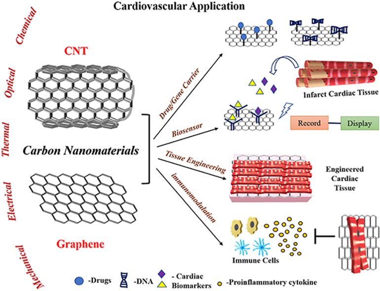
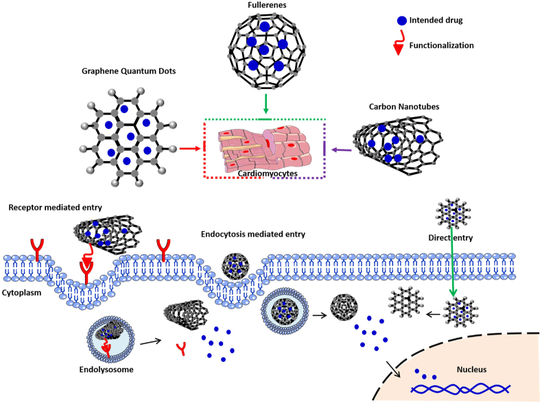
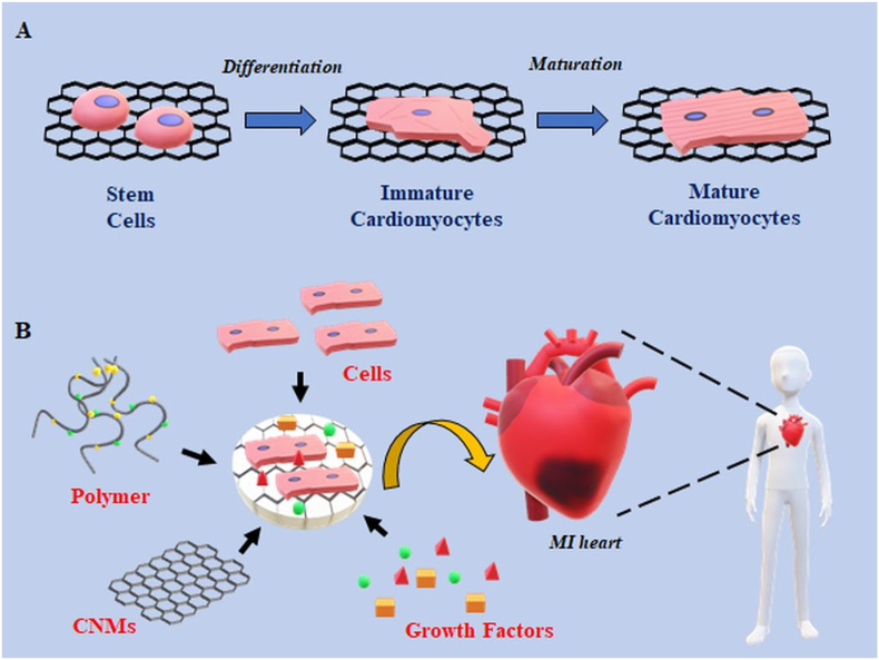
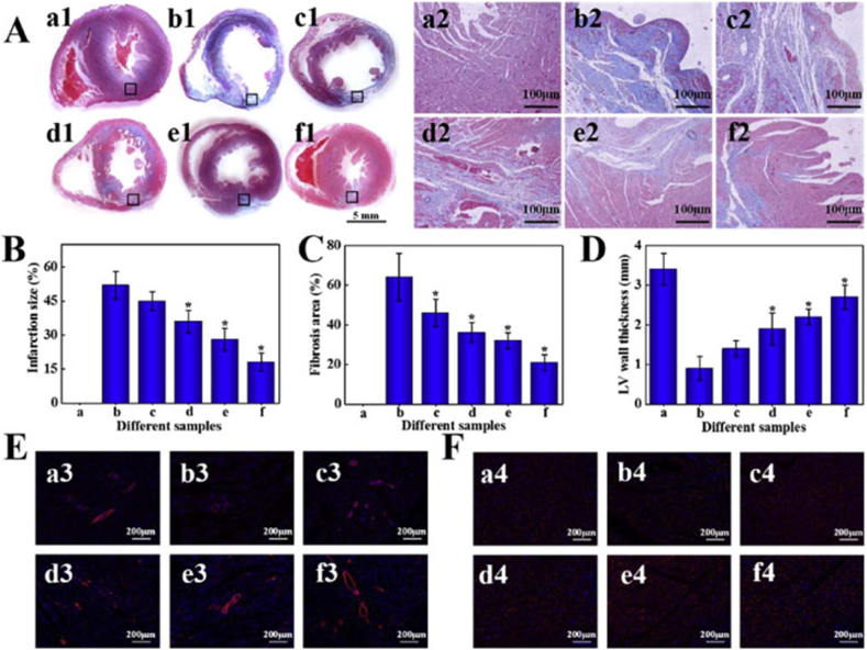
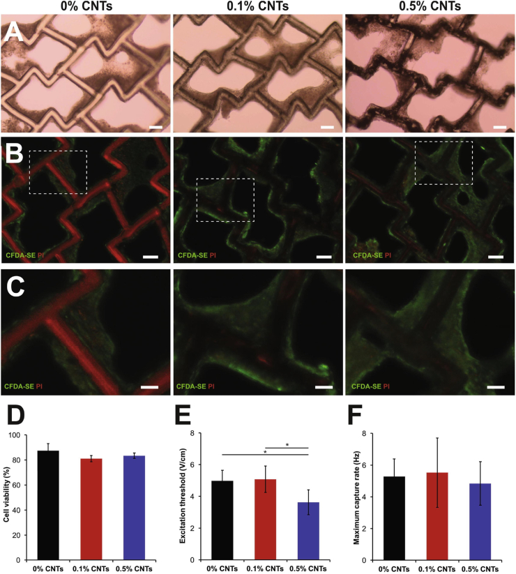
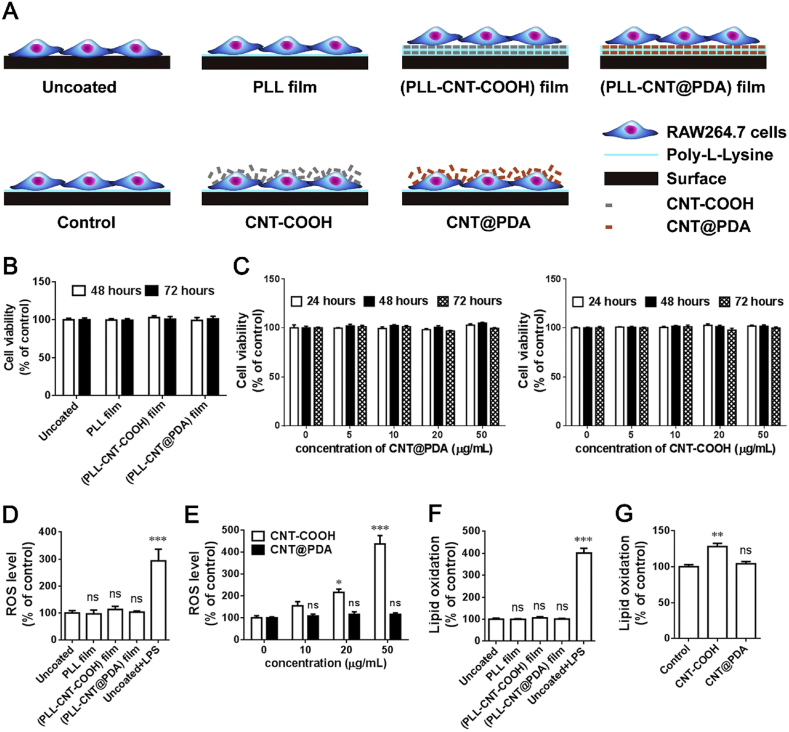
Cardiovascular diseases (CVDs) are the leading cause of death worldwide. Heart attack and stroke cause irreversible tissue damage. The currently available treatment options are limited to "damage-control" rather than tissue repair. The recent advances in nanomaterials have offered novel approaches to restore tissue function after injury. In particular, carbon nanomaterials (CNMs) have shown significant promise to bridge the gap in clinical translation of biomaterial based therapies. This family of carbon allotropes (including graphenes, carbon nanotubes and fullerenes) have unique physiochemical properties, including exceptional mechanical strength, electrical conductivity, chemical behaviour, thermal stability and optical properties. These intrinsic properties make CNMs ideal materials for use in cardiovascular theranostics. This review is focused on recent efforts in the diagnosis and treatment of heart diseases using graphenes and carbon nanotubes. The first section introduces currently available derivatives of graphenes and carbon nanotubes and discusses some of the key characteristics of these materials. The second section covers their application in drug delivery, biosensors, tissue engineering and immunomodulation with a focus on cardiovascular applications. The final section discusses current shortcomings and limitations of CNMs in cardiovascular applications and reviews ongoing efforts to address these concerns and to bring CNMs from bench to bedside.

Keywords: Biosensors; Carbon nanomaterials; Cardiac tissue engineering; Cardiovascular disease; Drug delivery; Immunomodulation.


Conflict of interest statement

Authors declare no conflict of interest.

Figures

Image 1
Graphical abstract
Fig. 1
Fig. 1
Schematic representation of different mechanistic approaches employed by CNMs as carriers to deliver drugs/gene into the cardiomyocytes.
Fig. 2
Fig. 2
Preparation of injectable hydrogel for acute myocardial infarction (AMI) therapy. (a) Schematic of stepwise formulation process of bioactive hydrogel and subsequent injection to treat damaged heart with acute myocardial infarction. (1) First, GO nanosheets are functionalized by amide bond with branched PEI to form cationic fGO. (2) fGO is then surface functionalized with anionic plasmids (DNAVEGF) to form fGO/DNAVEGF as shown in TEM images. (3) These bioactive hybrids are then suspended in prepolymer of GelMA hydrogel and UV cross-linked under optimized condition to form hydrogel (4) injectable fGO/DNAVEGF carrying GelMA hydrogel (GG′). (5) The latter is then intramyocardially injected in rat heart with AMI for local gene delivery of incorporated fGO/DNAVEGF nanocomplexes. (6) The hydrogel exhibits therapeutic effects by promoting myocardial vasculogenesis, which leads to reduced scar area and improved cardiac function. (b) Injectability of the developed GO carrying GelMA hydrogel. The viscosity of GO/GelMA nanocomposite hydrogels was monitored at different shear rates. At low shear rate, both fGO/GelMA and GelMA hydrogels had high viscosity. However, at higher shear rate, fGO/GelMA and GelMA hydrogels showed decreased viscosity. This suggests that both GelMA and fGO/GelMA were able to flow at higher shear rate and were easily injectable. The results also indicate that the addition of surface functionalized fGO to GelMA results in higher viscosity of fGO/GelMA at higher shear rate compared to GelMA. In other words, fGO reinforces the GelMA hydrogel network. Scale bar: 1 μm(reproduced with permission from Ref. [86], © American Chemical Society, 2020).
Fig. 3
Fig. 3
Application of CNMs in biosensors for diagnosis of CVDs. Model demonstrates the detection of cardiac biomarkers in blood of CVD patients. The sensors have biocatalysts such as DNA, enzymes, antibodies present on the surface which are used to detect specific cardiac biomarkers. The signals are then amplified and processed to display the value.
Fig. 4
Fig. 4
a) Schematic illustration of the PtNPs-decorated rGO FET biosensor with a custom-made microfilter for BNP detection. b). (A) Real-time electrical detection at different concentrations of BNP solution in PBS (12.5 nM, 25 nM, 50 nM, 100 nM, 200 nM, 400 nM, 800 nM). Inset: Real-time electrical detection at lower concentrations of BNP. (B) The response of the PtNPs-decorated rGO biosensors to BNP at different concentrations. Inset: The response of the biosensors to BNP at low concentration range (100 fM, 1 pM, 10 pM, 100 pM, 1 nM) (Reproduced with permission from Ref. [119].© Elsevier Ltd 2020).
Fig. 5
Fig. 5
Schematic illustration showing (A) the fabrication of the MIP sensor and (B) the binding detection. Sensor performance: (C) effects of interfering substances on the response of the cTnT-MIP sensors after the addition of cTnI, HSA, Glu, AA, Cr and UA in a solution containing 1.0 pg mL-1 cTnT and the different concentrations of cTnI, HSA, Glu, AA, Cr and UA were tested using the developed MIP sensor. (D) differential pulse voltammograms (DPVs) of the developed MIP sensor obtained with 0.0–40 pg mL−1 of cTnT in 0.10 mol l−1 PBS (pH 7.00). (E) plot of the change in current response (ΔI) versus concentration of cTnT of the MIP and NIP sensors fitted with the Langmuir isotherm using GraphPad Prism 8 and the inset showing the linear range (n = 3) (Reproduced with permission from Ref. [136].© Elsevier Ltd 2020).
Fig. 6
Fig. 6
Overview of CNMs in cardiac tissue engineering. A) Application of CNMs in differentiation and maturation of stem cells derived cardiomyocytes. CNMs serve as reinforcers in scaffold/hydrogels which promotes differentiation of stem cells and maturation of stem cells derived cardiomyocytes. B) Construction of CNMs-based cardiac constructs for treatment in MI. CNMs provide mechanical strength and electrical conductivity to a cardiac patch which is comprised of cells, polymers and growth factors for effective myocardial repair during MI.
Fig. 7
Fig. 7
Cardiac repair by different treatments after 28 days (a, a1-a4: Sham; b, b1-b4: PBS; c, c1-c4: PEG-MEL/HA-SH; d, d1-d4: PEG-MEL/HA-SH/GO; e, e1-e4: PEG-MEL/HA-SH/ADSCs; f, f1-f4: PEG-MEL/HA-SH/GO/ADSCs). (A) Masson's trichrome staining for collagen (blue) and muscle (red); a2-f2 is magnification of the corresponding black box labelled in a1-f1. (B) Infarction size. (C) Fibrosis area. (D) LV wall thickness. (E) Immunofluorescence staining for α-SMA. (F) Immunofluorescence staining for Cx43. * indicates a significant difference between the experimental group and PBS treated group (Reproduced with permission from Ref. [154].© Elsevier Ltd 2020).
Fig. 8
Fig. 8
Composite scaffolds in cardiac tissue engineering. (A–C) Images of rat cardiac tissue constructs with 124 polymer-CNT scaffolds containing 0.1% and 0.5% CNTs compared to pure polymer scaffolds. (A) Bright field images of polymers seeded with rat CMs at day 7 of culture demonstrate tissue compaction around scaffold struts (Scale bars: 100 μm). (B) Tissues were imaged for viability with live/dead staining assay (Live cells: green, dead cells: red), where the scaffold exhibits autofluorescence in the red channel (Scale bars: 100 μm). (C) High magnification excerpts demonstrate the wrapping of viable cells around scaffold struts (Scale bars: 50 μm). (D) The quantification of live-dead assay images present no difference in cell viability among material groups. (E and F) Comparison of excitation threshold (E) and maximum capture rate (F) suggest improved tissue properties with 0.5% CNTs in polymer scaffolds in comparison to pure polymer controls (∗p < 0.05) (Reproduced with permission from Ref. [167].© Elsevier Ltd 2020).
Fig. 9
Fig. 9
(A). Schematic diagram of experimental design to evaluate material biocompatibility with RAW264.7 macrophages. The cells were cultured on (PLL-CNT-COOH) film or (PLL-CNT@PDA) film, and then examined for cellular functions. The cells grown on the surface without modification (Uncoated) and PLL-coated surface (PLL film) were applied as controls. RAW264.7 cells were also directly treated with dispersed CNTs (CNT-COOH or CNT@PDA), using untreated cells as control. B, The cells were seeded onto different films for 48 and 72 h. Cell viability examined by the CCK-8 assay was comparable among these groups. C, The cells were treated with CNT@PDA or CNT-COOH at different concentrations for 24, 48 and 72 h. No viability change was observed. D, ROS levels were detected by the DCFH-DA assay. No change in ROS levels was found in RAW264.7 cells following a 72-h culture on the specified films. E, A 24 h treatment of CNT-COOH increased ROS levels in a concentration-dependent manner, whereas CNT@PDA had no significant effect even at a concentration of 50 μg/mL. F, Treatments with none of the films for 72 h affected lipid oxidation, as determined by the MDA assay. G, CNT-COOH, but not CNT@PDA treatment for 24 h raised cellular lipid oxidation. n = 4–6; *P < 0.05, **P < 0.01, ***P < 0.001 versus uncoated or specified controls by one-way ANOVA; ns: not significant (Reproduced with permission from Ref. [190].© Elsevier Ltd 2020).

References

    1. Flora G.D., Nayak M.K. A brief review of cardiovascular diseases, associated risk factors and current treatment regimes. Curr. Pharmaceut. Des. 2019;25:4063–4084. doi: 10.2174/1381612825666190925163827. - DOI - PubMed
    1. Barquera S., Pedroza-Tobías A., Medina C., Hernández-Barrera L., Bibbins-Domingo K., Lozano R., Moran A.E. Global Overview of the epidemiology of atherosclerotic cardiovascular disease. Arch. Med. Res. 2015;46:328–338. doi: 10.1016/j.arcmed.2015.06.006. - DOI - PubMed
    1. Nabel E.G. Cardiovascular disease. N. Engl. J. Med. 2003;349:60–72. doi: 10.1056/NEJMra035098. - DOI - PubMed
    1. Burke A.P., Virmani R. Pathophysiology of acute myocardial infarction. Med. Clin. 2007;91:553–572. doi: 10.1016/j.mcna.2007.03.005. ix. - DOI - PubMed
    1. Frangogiannis N.G. Comprehensive Physiology. American Cancer Society; 2015. Pathophysiology of myocardial infarction; pp. 1841–1875. - DOI - PubMed

LinkOut - more resources