. 2021 Jan 26;6(8):2372-2389.
doi: 10.1016/j.bioactmat.2021.01.017. eCollection 2021 Aug.

Cartilage-targeting peptide-modified dual-drug delivery nanoplatform with NIR laser response for osteoarthritis therapy

Affiliations

Cartilage-targeting peptide-modified dual-drug delivery nanoplatform with NIR laser response for osteoarthritis therapy

Song Xue et al. Bioact Mater. .

Abstract

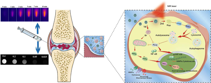
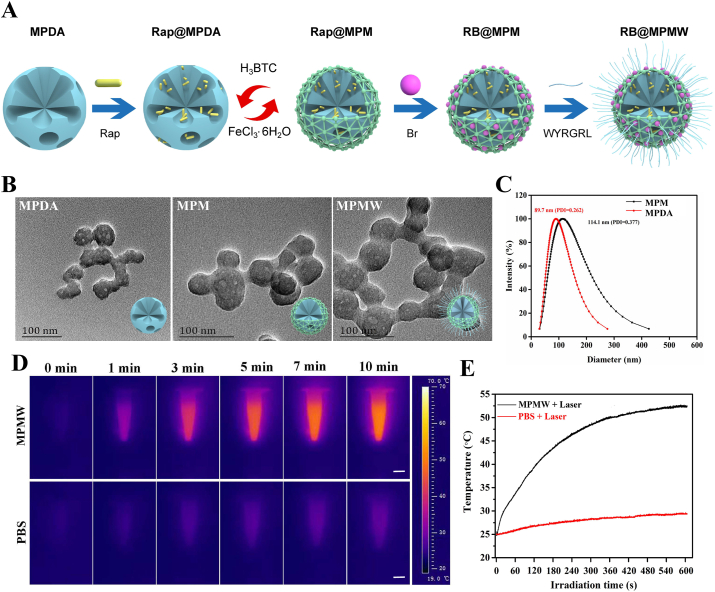
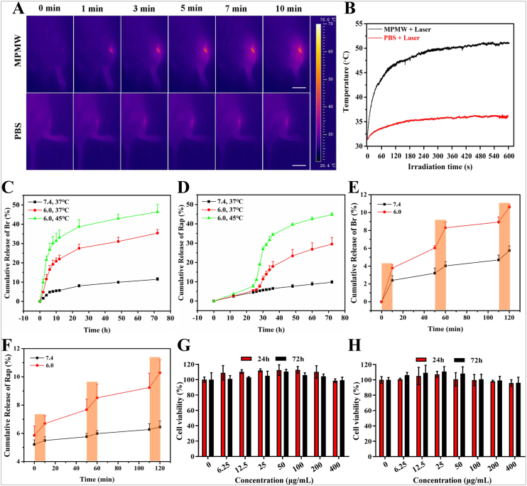
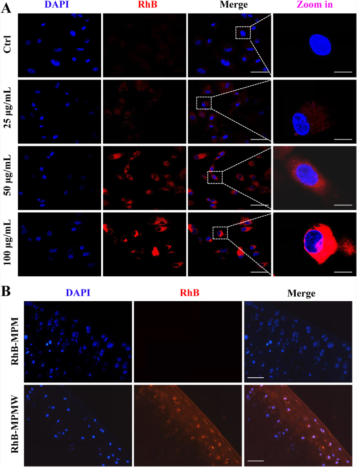
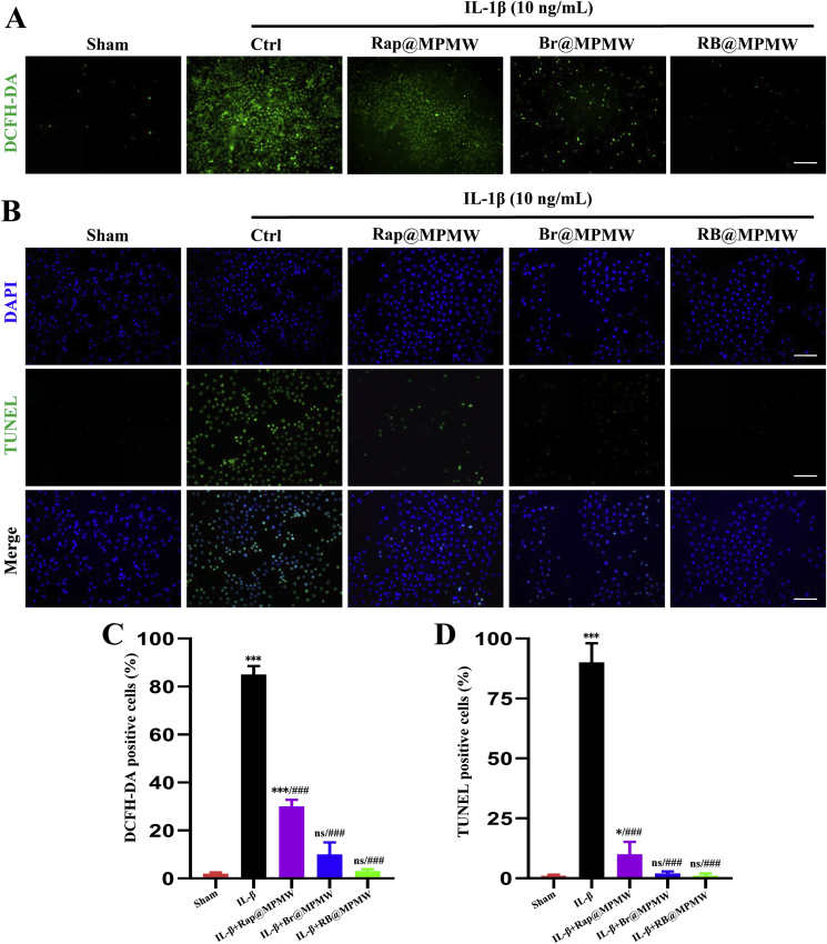
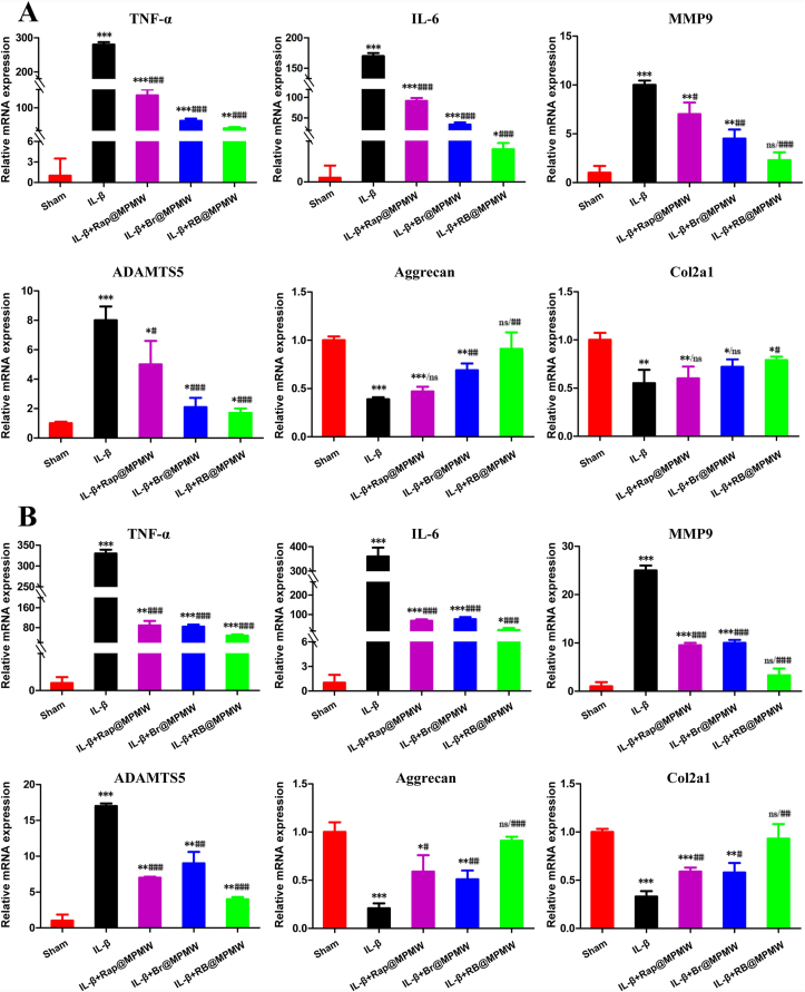
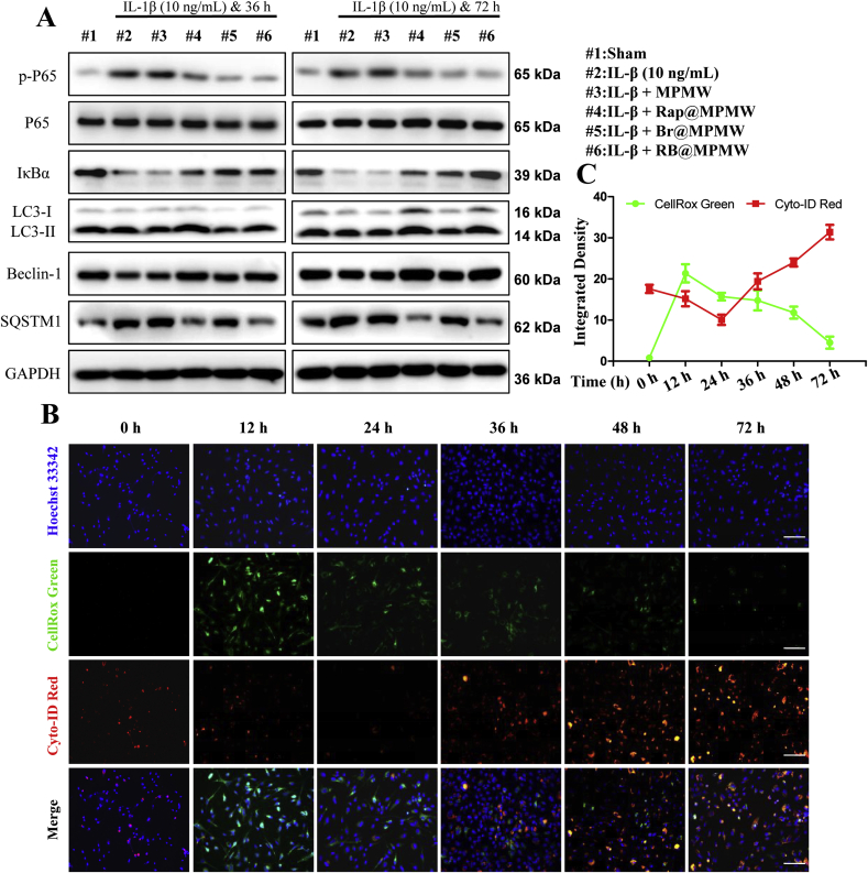
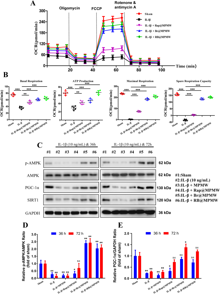
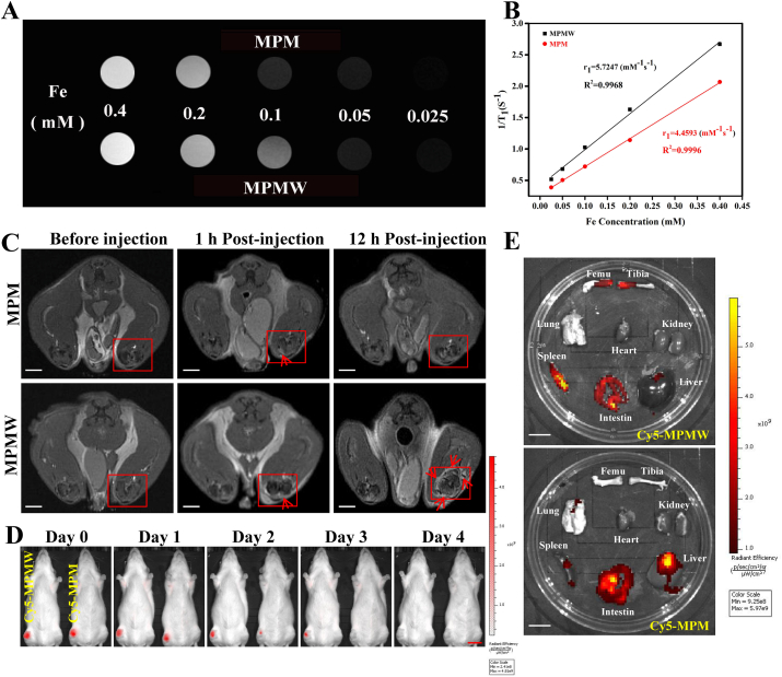
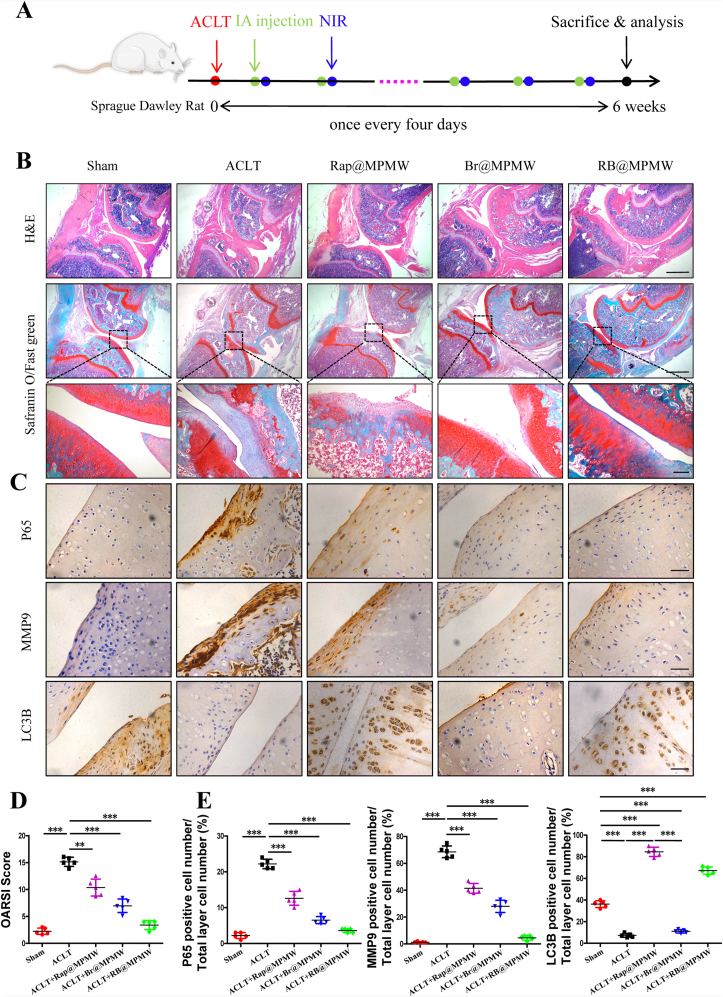
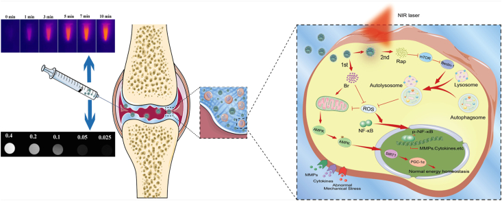
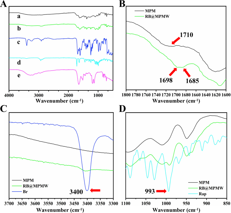
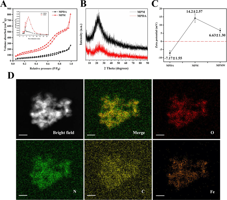
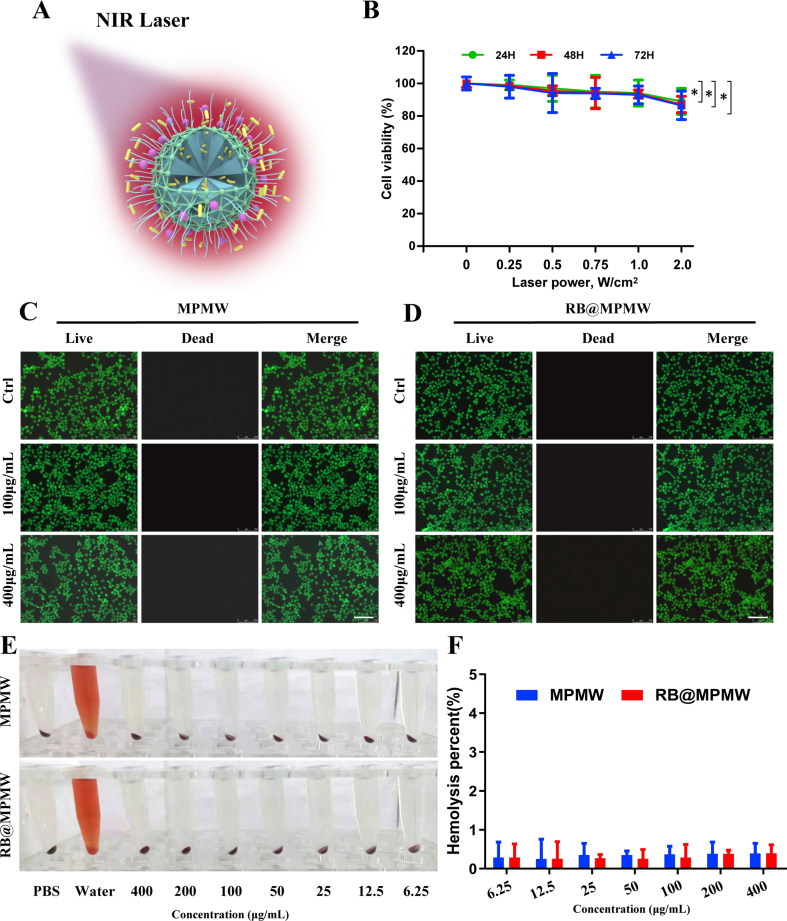
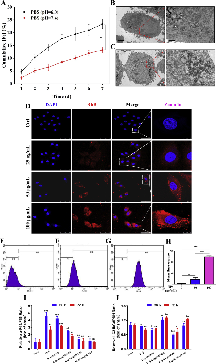
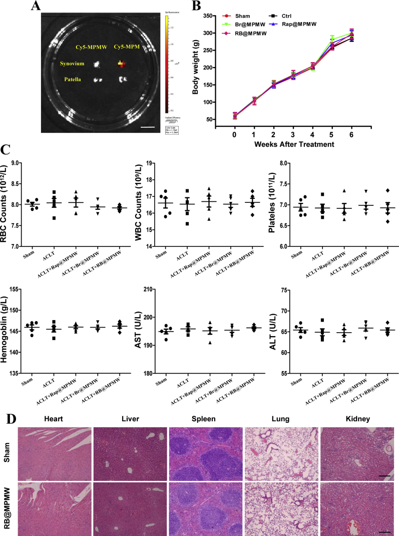
Cartilage-targeting delivery of therapeutic agents is still an effective strategy for osteoarthritis (OA) therapy. Recently, scavenging for reactive oxygen species (ROS) and activating autophagy have been increasingly reported to treat OA effectively. In this study, we designed, for the first time, a dual-drug delivery system based on metal organic framework (MOF)-decorated mesoporous polydopamine (MPDA) which composed of rapamycin (Rap) loaded into the mesopores and bilirubin (Br) loaded onto the shell of MOF. The collagen II-targeting peptide (WYRGRL) was then conjugated on the surface of above nanocarrier to develop a cartilage-targeting dual-drug delivery nanoplatform (RB@MPMW). Our results indicated the sequential release of two agents from RB@MPMW could be achieved via near-infrared (NIR) laser irritation. Briefly, the rapid release of Br from the MOF shell exhibited excellent ROS scavenging ability and anti-apoptosis effects, however responsively reduced autophagy activity, to a certain extent. Meanwhile, following the NIR irradiation, Rap was rapidly released from MPDA core and further enhanced autophagy activation and chondrocyte protection. RB@MPMW continuously phosphorylated AMPK and further rescued mitochondrial energy metabolism of chondrocytes following IL-1β stimulation via activating SIRT1-PGC-1α signaling pathway. Additionally, the cartilage-targeting property of peptide-modified nanocarrier could be monitored via Magnetic Resonance (MR) and IVIS imaging. More significantly, RB@MPMW effectively delayed cartilage degeneration in ACLT rat model. Overall, our findings indicated that the as-prepared dual-drug delivery nanoplatform exerted potent anti-inflammation and anti-apoptotic effects, rescued energy metabolism of chondrocytes in vitro and prevented cartilage degeneration in vivo, which thereby showed positive performance for OA therapy.

Keywords: Autophagy; Energy metabolism; Osteoarthritis; Oxidative stress; Targeting therapy.


Conflict of interest statement

All authors declared that no conflict interest existed.

Figures

Image 1
Graphical abstract
Fig. 1
Fig. 1
(A) The schematic diagram of fabrication of RB@MPMW. (B) TEM images of MPDA, MPM and MPMW, respectively. (C) DLS of MPDA & MPM in deionized water. (D) Photothermal performance of MPMW (100 μg/mL) in aqueous solution under NIR irradiation (808 nm, 1 W/cm2) at different time points (scale bar: 0.3 cm). (E) The temperature changes at indicated time in vitro with or without MPMW.
Fig. 2
Fig. 2
(A) Photothermal performance of MPMW (100 μg/mL) in the knee joint under NIR laser irradiation (808 nm, 1 W/cm2) at different time points (scale bar: 2 cm). (B) The temperature changes at indicated time in vivo with or without MPMW. (C,D) The cumulative release of Br and Rap from the BR@MPMW at different pH (7.4 or 6.0) and temperature (37 °C or 45 °C), respectively. (E,F) NIR laser-triggered release of dual agents under NIR laser irradiation at different pH (7.4 or 6.0) and different time points. The cumulative release of Rap was start to assessed after incubation for 24 h at indicated conditions. (G,H) Cell viability assay of ATDC5 cells incubated with (G) MPMW and (H) RB@MPMW at different concentrations (0, 6.25, 12.5, 25, 50, 100, 200 and 400 μg/mL) under NIR laser irridation for 10 min every 12 h.
Fig. 3
Fig. 3
(A) CLSM images of ATDC5 cells incubated with different concentrations of RhB-MPMW for 4 h (scale bar: 45 μm (before zoom in); 15 μm (after zoom in)). (B) The histological fluorescence images of articular cartilage pretreated with RhB-MPM (upper) & RhB-MPMW (lower) for two days (scale bar: 45 μm).
Fig. 4
Fig. 4
(A) ROS measurement of ATDC5 cells after different samples treatment and subsequent IL-1β (10 ng/mL) stimulation for 72 h (scale bar: 45 μm). (B) Anti-apoptotic effects on ATDC5 cells of different samples after stimulation with IL-1β (10 ng/mL) for 72 h (scale bar: 45 μm). (C,D) The percent of DCFH-DA positive cells and TUNEL positive cells, respectively. * indicates p < 0.05; ***, ### indicate p < 0.001; ns indicates not significant. * indicates comparisons with the sham group and # indicates comparisons with the IL-1β treatment group.
Fig. 5
Fig. 5
The expression of OA-related genes (TNF-α, IL-6, MMP9, ADAMTS5, Aggrecan and Col2a1) after (A) 36 h and (B) 72 h treatment with different samples and further exposure to IL-1β (10 ng/mL). Data are shown as the means ± SD of triplicate independent experiments. *, # indicate p < 0.05; **, ## indicate p < 0.01; ***, ### indicate p < 0.001; ns indicates not significant. * indicates comparisons with the sham group and # indicates comparisons with the IL-1β treatment group.
Fig. 6
Fig. 6
(A) Some key proteins expression in autophagy and NF-κB signal pathway. (B) Fluorescence images of ATDC5 cells stained with Cyto-ID Red and CellRox Green after treated with RB@MPMW (50 μg/mL), IL-1β (10 ng/mL) and NIR laser (808 nm, 1 W/cm2) for different incubation time (scale bar: 25 μm). (C) Quantitative analysis of dynamic changes of autophagy activity and ROS via calculating average intensity by ImageJ software.
Fig. 7
Fig. 7
(A) OCR of ATDC5 cells after different treatments. (B) Some parameters were shown based on the OCR analysis, including basal respiration, ATP production, maximal respiration and spare respiration capacity. (C) The effects of various treatments on the AMPK-SIRT1-PGC-1α signaling pathway were determined. (D,E) The relative expression of p-AMPK/AMPK and PGC-1α/GAPDH among various groups (compared with the sham group). * indicates p < 0.05; ** indicates p < 0.01; *** indicates p < 0.001; ns indicates not significant.
Fig. 8
Fig. 8
(A) The T1-weight MR images of MPM & MPMW with different Fe concentrations (0.025, 0.05, 0.1, 0.2 and 0.4 mM) in vitro. (B) The respective r1 relaxivity curves of MPM & MPMW. (C) The T1-weight MR images of ACLT rat model intra-articularly treated with MPM & MPMW (50 μg/mL, 20 μL) for indicated time points (scale bar: 0.5 cm). The red box shows the image of the right knee joint before and after the injection of MPM & MPMW, while the arrow mainly shows the signal enhancement area of the cartilage before and after the injection. (D) The IVIS images of ACLT rat model after IA injection of MPM & MPMW (50 μg/mL, 20 μL) for indicated time points (scale bar: 2 cm). (E) The IVIS images of some important organs to show the retention of nanoparticles at 48 h after IA injection (scale bar: 2 cm).
Fig. 9
Fig. 9
(A) Treatment schedule. (B) H&E staining and Safranin O/Fast green staining of the articular cartilage after 6 weeks treatment with different samples (scale bar: top and center, 450 μm; bottom, 150 μm). (C) Immunohistochemisy analysis of the articular cartilage after 6 weeks treatment with different samples (n = 5, scale bar: 45 μm). (D) The overall OARSI scores of the articular cartilage. (E) Relative expression of P65, MMP9 and LC3B for each group. ** indicate p < 0.01; *** indicate p < 0.001.
Fig. 10
Fig. 10
The mechanism of dual-drug delivery nanoplatform with cartilage-targeting effect and NIR laser response for OA therapy.
figs1
figs1
figs2
figs2
figs3
figs3
figs4
figs4
figs5
figs5
figs6
figs6

References

    1. Kloppenburg M., Berenbaum F. Osteoarthritis year in review 2019: epidemiology and therapy. Osteoarthritis Cartilage. 2020;28:242–248. - PubMed
    1. Nasiri N., Hosseini S., Alini M., Khademhosseini A., Baghaban Eslaminejad M. Targeted cell delivery for articular cartilage regeneration and osteoarthritis treatment, Drug Discov. Today Off. 2019;24:2212–2224. - PubMed
    1. Fraenkel L., Buta E., Suter L., Dubreuil M., Levy C., Najem C., Brennan M., Corn B., Kerns R., Goulet J. Nonsteroidal anti-inflammatory drugs vs cognitive behavioral therapy for arthritis pain: a randomized withdrawal trial. JAMA Intern. Med. 2020 - PMC - PubMed
    1. da Costa B.R., Reichenbach S., Keller N., Nartey L., Wandel S., Juni P., Trelle S. Effectiveness of non-steroidal anti-inflammatory drugs for the treatment of pain in knee and hip osteoarthritis: a network meta-analysis. Lancet. 2017;390:e21–e33. - PubMed
    1. Lepetsos P., Papavassiliou A.G. ROS/oxidative stress signaling in osteoarthritis. Biochim. Biophys. Acta. 2016;1862:576–591. - PubMed

LinkOut - more resources