. 2021 Feb 2;6(8):2479-2490.
doi: 10.1016/j.bioactmat.2021.01.009. eCollection 2021 Aug.

Milk exosomes: Nature's abundant nanoplatform for theranostic applications

Affiliations

Milk exosomes: Nature's abundant nanoplatform for theranostic applications

Benilde Adriano et al. Bioact Mater. .

Abstract

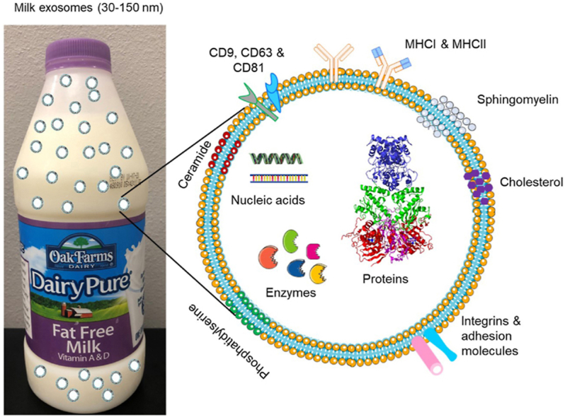
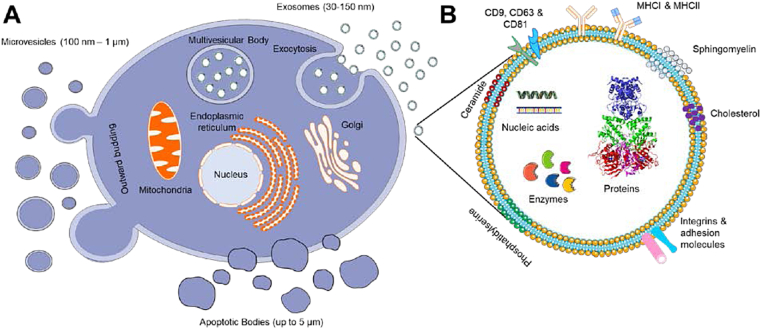
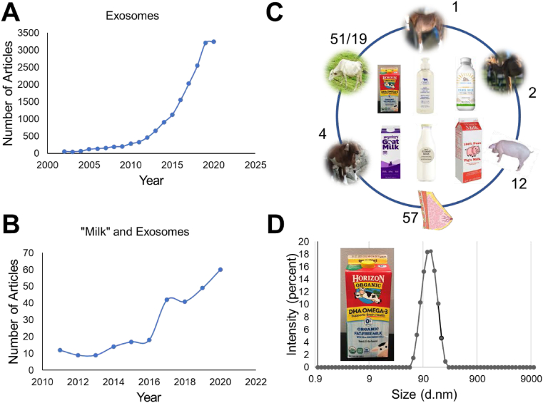
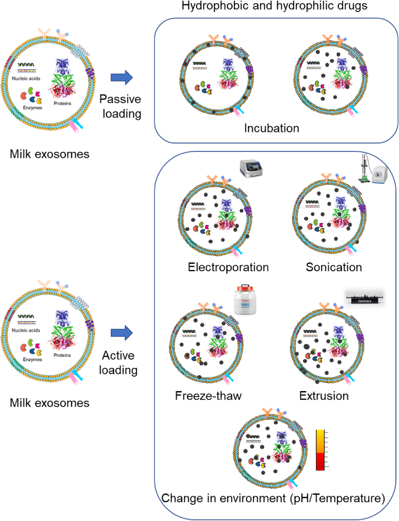
Exosomes are a unique subpopulation of naturally occurring extracellular vesicles which are smaller intracellular membrane nanoparticle vesicles. Exosomes have proven to be excellent nanocarriers for carrying lipids, proteins, mRNAs, non-coding RNAs, and DNAs, and disseminating long-distance intercellular communications in various biological processes. Among various cell-line or biological fluid derived exosomes, milk exosomes are abundant in nature and exhibit many nanocarrier characteristics favorable for theranostic applications. To be an effective delivery carrier for their clinical translation, exosomes must inbuilt loading, release, targeting, and imaging/tracking characteristics. Considering the unmet gaps of milk exosomes in theranostic technology it is essential to focus the current review on drug delivery and imaging applications. This review delineates the efficiency of exosomes to load therapeutic or imaging agents and their successful delivery approaches. It is emphasized on possible modifications of exosomes towards increasing the stability and delivery of agents. This article also summarizes the specific applications and the process of developing milk exosomes as a future pharmaceutical drug delivery vehicle.

Keywords: Drug delivery; Extracellular vesicles; Imaging agents; Milk exosomes; Theranostic applications.


Conflict of interest statement

The authors declare that they have no known competing financial interests or personal relationships that could have appeared to influence the work reported in this paper.

Figures

Image 1
Graphical abstract
Fig. 1
Fig. 1
A) A schematic representation of cellular vesicle production (exosomes, microvesicles, and apoptotic bodies) from cells including milk. Distinct variation in the biogenesis and excretion from cell to yield different size range excretes. B) An illustration of exosome structure with rich source for protein, nucleic acid, enzyme, lipids, and cargos.
Fig. 2
Fig. 2
A-C) Peer-reviewed scientific reports and review articles covering various aspects of exosomes recorded using PubMed website https://www.ncbi.nlm.nih.gov/. All data was retrieved on Oct 12, 2020. A) Number of articles found that are published in the literature in each year since 2001 to 2020. B) Number of articles noticed in each year with key word of “Milk” and exosomes, C) Articles found in search with key words of “human milk” and exosome, “bovine/cow milk” and exosome, “horse milk” and exosome, “camel milk” and exosome, “goat milk” and exosome, and “porcine milk” and exosome. D) An example of size distribution of bovine milk exosomes derived from fat free Horizon organic milk using acetic acid precipitation method. It shows a uniform particle size of ~100 nm.
Fig. 3
Fig. 3
The schematic illustration of drug loading or encapsulation in exosomes. A) A simple post representation incubation strategy for passive diffusion into exosome structures. B) An illustrative presentation of stimuli or active drug loading methods in exosomes with the help of electroporation (transfection), sonication, freeze-thaw, extrusion, and temperature/pH gradiation.
Fig. 4
Fig. 4
Different strategies explored for engineering of exosome surface with imaging probe (radioisotope/radiotracer, magnetic nanoparticles, gold, quantum dots, and labelling fluorescent probes), targeting moieties (monoclonal/polyclonal antibodies, aptamer, fragmented antibodies, and other biomacromolecules), conjugating small, biological, macro-/bio-macromolecules (reporter agents and other molecules), and hybrid nanosystems. (For interpretation of the references to colour in this figure legend, the reader is referred to the Web version of this article.)

References

    1. Johnstone R.M., Adam M., Hammond J.R., Orr L., Turbide C. Vesicle formation during reticulocyte maturation. Association of plasma membrane activities with released vesicles (exosomes) J. Biol. Chem. 1987;262(19):9412–9420. - PubMed
    1. Johnstone R.M. Revisiting the road to the discovery of exosomes. Blood Cell Mol. Dis. 2005;34(3):214–219. - PubMed
    1. Johnstone R.M., Mathew A., Mason A.B., Teng K. Exosome formation during maturation of mammalian and avian reticulocytes: evidence that exosome release is a major route for externalization of obsolete membrane. Proteins. 1991;147(1):27–36. - PubMed
    1. Lázaro-Ibáñez E., Sanz-Garcia A., Visakorpi T., Escobedo-Lucea C., Siljander P., Ayuso-Sacido A., Yliperttula M. Different gDNA content in the subpopulations of prostate cancer extracellular vesicles: apoptotic bodies, microvesicles, and exosomes. Prostate. 2014;74(14):1379–1390. - PMC - PubMed
    1. Akers J.C., Gonda D., Kim R., Carter B.S., Chen C.C. Biogenesis of extracellular vesicles (EV): exosomes, microvesicles, retrovirus-like vesicles, and apoptotic bodies. Journal of neuro-oncology. 2013;113(1):1–11. - PMC - PubMed