. 2021 Jan 25;6(4):3120-3129.
doi: 10.1021/acsomega.0c05516. eCollection 2021 Feb 2.

Usage of Quantum Chemical Methods to Understand the Formation of Concomitant Polymorphs of Acetyl 2-(N-(2-Fluorophenyl)imino)coumarin-3-carboxamide

Affiliations

Usage of Quantum Chemical Methods to Understand the Formation of Concomitant Polymorphs of Acetyl 2-(N-(2-Fluorophenyl)imino)coumarin-3-carboxamide

Svitlana V Shishkina et al. ACS Omega. .

Abstract

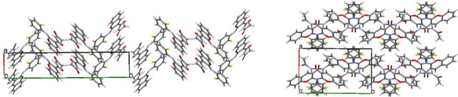
Crystallization of concomitant polymorphs is a very intriguing process that is difficult to be studied experimentally. A comprehensive study of two polymorphic modifications of acetyl 2-(N-(2-fluorophenyl)imino)coumarin-3-carboxamide using quantum chemical methods has revealed molecular and crystal structure dependence on crystallization conditions. Fast crystallization associated with a kinetically controlled process results in the formation of a columnar structure with a nonequilibrium molecular conformation and more isotropic topology of interaction energies between molecules. Slow crystallization may be considered as a thermodynamically controlled process and leads to the formation of a layered crystal structure with the conformation of the molecule corresponding to local minima and anisotropic topology of interaction energies. Fast crystallization results in the formation of a lot of weak intermolecular interactions, while slow crystallization leads to the formation of small amounts of stronger interactions.


Conflict of interest statement

The authors declare no competing financial interest.

Figures

Scheme 1
Scheme 1. Acetyl 2-(N-(2-Fluorophenyl)imino)coumarin-3-carboxamide
Figure 1
Figure 1
2-(N-(2-Fluorophenyl)imino)coumarin-3-carboxamide molecular structures in polymorphic modifications I (on the left) and II (on the right) according to the X-ray diffraction data.
Figure 2
Figure 2
Molecular structure of two equilibrium states (along the edges) and transition state (in the middle) according to the quantum chemical calculations.
Figure 3
Figure 3
Energy profile of the rotation around the N1–C10 bond in unsubstituted and ortho-fluorosubstituted molecules in vacuum (on the left) and taking into account the polarizing environment within the PCM model (solvent = isopropanol) according to the quantum chemical calculations by the m06-2x/cc-pVTZ method.
Figure 4
Figure 4
Intermolecular hydrogen bonds in polymorphic structure I.
Figure 5
Figure 5
Stacking interactions in crystals I (on the left) and II (on the right).
Figure 6
Figure 6
Crystal structures of polymorphic modification I (on the left) and II (on the right).
Figure 7
Figure 7
Hirshfeld surfaces with the mapped dnorm property for molecules in structures I (on the left) and II (on the right) projected and transparent to show the conformation of the molecules.
Figure 8
Figure 8
2D Hirshfeld fingerprint plots for structures I (on the left) and II (in the middle). Relative contribution of different types of intermolecular interactions to the total Hirshfeld surface area (in %) is shown as the histogram (on the right).
Figure 9
Figure 9
Column along the a crystallographic direction as the primary BSM shown as packing of molecules (a) and energy-vector diagrams (b) (projection along the b crystallographic direction) and packing of the columns, in terms of energy-vector diagrams (c) (projection along the a crystallographic direction) in structure I. The double column is highlighted blue.
Figure 10
Figure 10
Layer parallel to the bc crystallographic plane, projection along the a crystallographic direction (on the left) and packing of the layer, projection along the c crystallographic direction (on the right) in structure II. The layer is highlighted in red.
Figure 11
Figure 11
Stacked dimers with the strongest interaction energy in structure II.
Figure 12
Figure 12
Needle-like crystals of polymorph I (on the left) and prismatic crystals of polymorph II (on the right).

References

    1. Coumarins: Biology, Applications, and Mode of Action; O’Kennedy R.; Thomas R. D., Eds.; Wiley: Chichester, U.K., 1997.
    1. Estévez-Braun A.; González A. G. Coumarins. Nat. Prod. Rep. 1997, 14, 465–475. 10.1039/np9971400465. - DOI - PubMed
    1. Murray R. D. H. Coumarins. Nat. Prod. Rep. 1995, 12, 477–505. 10.1039/np9951200477. - DOI
    1. Guo D.; Chen T.; Ye D.; Xu J.; Jiang H.; Chen K.; Wang H.; Liu H. Cell-permeable iminocoumarine-based fluorescent dyes for mitochondria. Org. Lett. 2011, 13, 2884–2887. 10.1021/ol200908r. - DOI - PubMed
    1. Sekar N. Coumarin dyes in laser technology. Colourage 2003, 50, 55–56.

LinkOut - more resources