. 2021 Jan 13;24(2):102058.
doi: 10.1016/j.isci.2021.102058. eCollection 2021 Feb 19.

Isoform-selective decrease of glycogen synthase kinase-3-beta (GSK-3β) reduces synaptic tau phosphorylation, transcellular spreading, and aggregation

Affiliations

Isoform-selective decrease of glycogen synthase kinase-3-beta (GSK-3β) reduces synaptic tau phosphorylation, transcellular spreading, and aggregation

Ana Claudia Amaral et al. iScience. .

Abstract

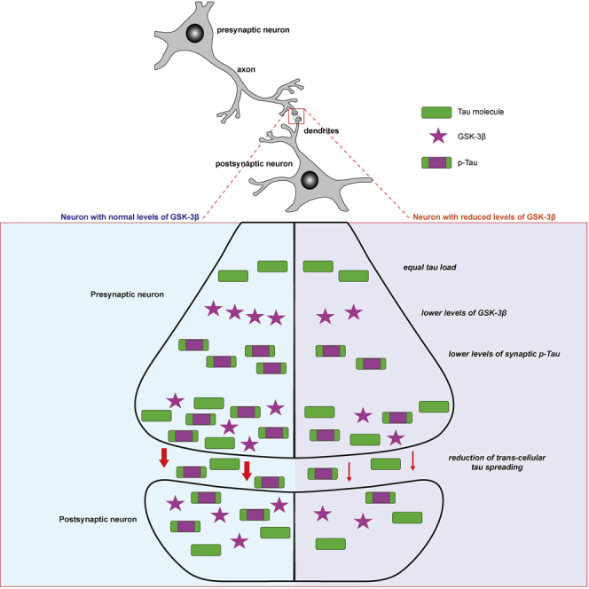
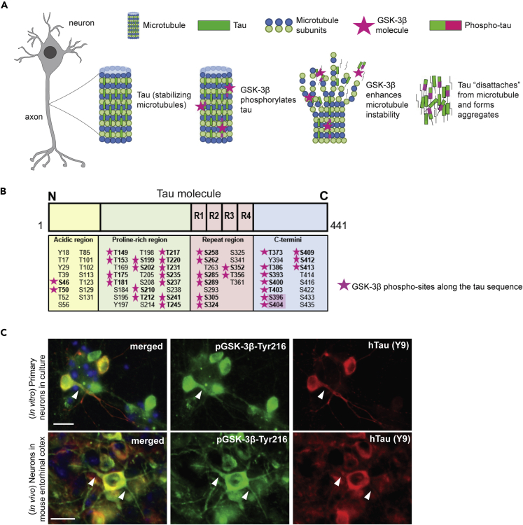
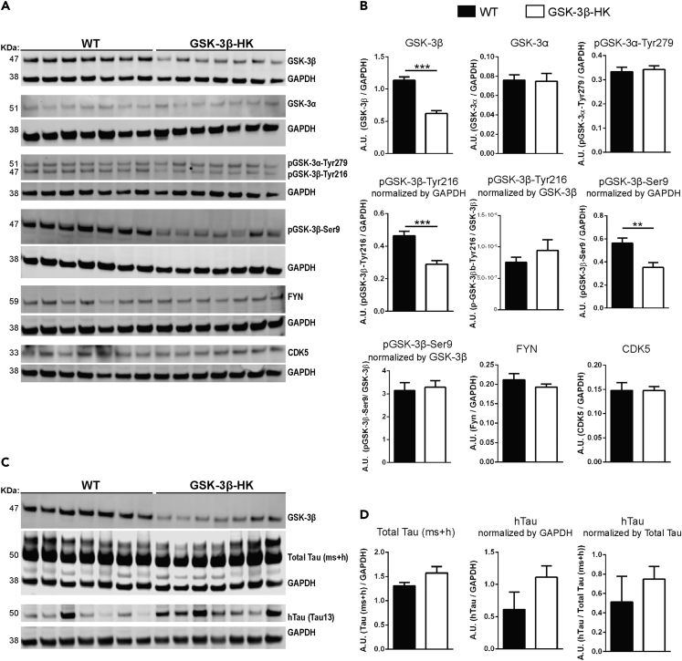
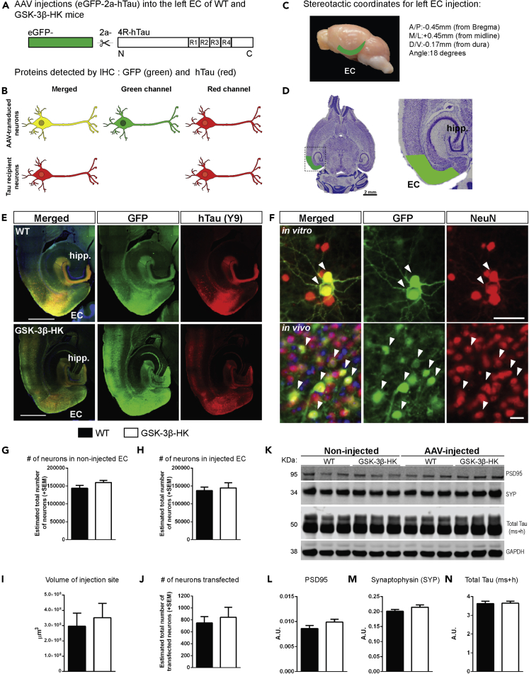
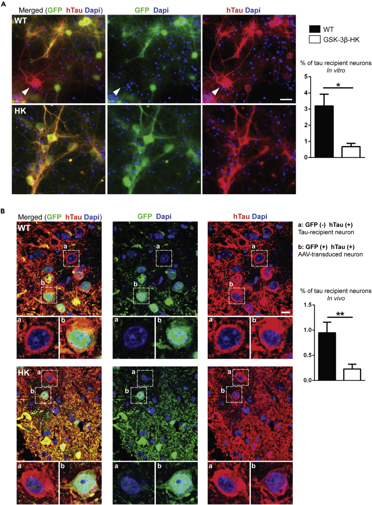
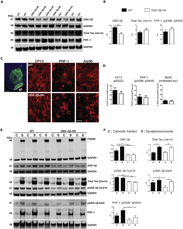
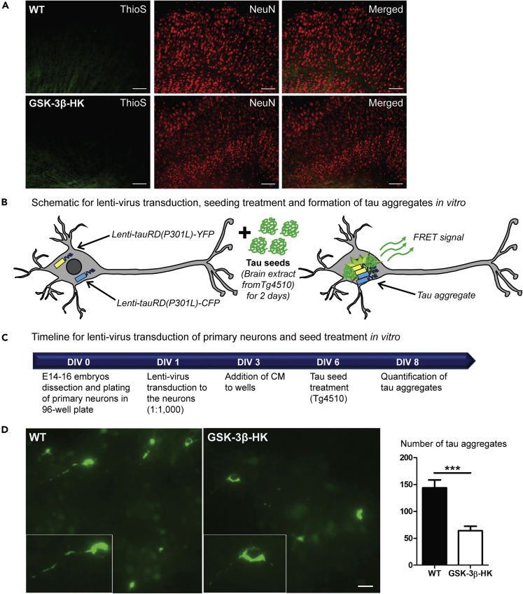
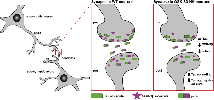
It has been suggested that aberrant activation of glycogen synthase kinase-3-beta (GSK-3β) can trigger abnormal tau hyperphosphorylation and aggregation, which ultimately leads to neuronal/synaptic damage and impaired cognition in Alzheimer disease (AD). We examined if isoform-selective partial reduction of GSK-3β can decrease pathological tau changes, including hyperphosphorylation, aggregation, and spreading, in mice with localized human wild-type tau (hTau) expression in the brain. We used adeno-associated viruses (AAVs) to express hTau locally in the entorhinal cortex of wild-type and GSK-3β hemi-knockout (GSK-3β-HK) mice. GSK-3β-HK mice had significantly less accumulation of hyperphosphorylated tau in synapses and showed a significant decrease of tau protein spread between neurons. In primary neuronal cultures from GSK-3β-HK mice, the aggregation of exogenous FTD-mutant tau was also significantly reduced. These results show that a partial decrease of GSK-3β significantly represses tau-initiated neurodegenerative changes in the brain, and therefore is a promising therapeutic target for AD and other tauopathies.

Keywords: Biological Sciences; Cellular Neuroscience; Neuroscience.


Conflict of interest statement

Teresa Gómez-Isla participated as speaker in an Eli Lilly and Company-sponsored educational symposium and serves in an Eli Lilly Data Monitoring Committee. All other authors declare no competing interests.

Figures

None
Graphical abstract
Figure 1
Figure 1
Tau phosphorylation by GSK-3β (A) Tau binds and stabilizes axonal microtubules in the brain, a process regulated by phosphorylation. Hyperphosphorylation of tau by different kinases, including GSK-3β, is associated with tau aggregation in AD. (B) GSK-3β phosphorylates tau at several epitopes (pink stars) across the tau sequence. Two phospho-sites targeted by GSK-3β, S396 and S404 (highlighted), are commonly hyperphosphorylated in AD and are found in neurofibrillary tangles. R1 to R4 refer to the four sequence repeats in the microtubule binding domain of tau. (C) Immunostaining of primary neurons from WT embryos after treatment with hTau (AAV added to culture media) and brain sections from an AAV-injected WT mouse showed that active GSK-3β (GSK-3β phosphorylated at Thr216) co-localizes with hTau (Y9) in the cytosol of neurons and in some neurites. Scale bars: 20 μm (C).
Figure 2
Figure 2
GSK-3β-HK mice model used in this study (A) Representative images of western blots using EC lysates from WT and GSK-3β-HK mice probed for kinases known to phosphorylate tau. (B) Quantification of western blots. Results showed a significant overall reduction of 45% of GSK-3β and 40% of pGSK-3β (active and inhibited forms, -Tyr216 and -Ser9, respectively) in GSK-3β-HK compared with WT mice. No differences were found in GSK-3α, pGSK-3α-Tyr279 (active GSK-3α), FYN, and CDK5 between WT and GSK-3β-HK mice. Data are presented as mean ± SEM, N = 14 mice: 7WT, 7HK. Two-tailed Student's t test, ∗∗p < 0.01, ∗∗∗p < 0.001. (C) Representative images of western blots probed for hTau and Total Tau (mouse + human tau (ms + h)) in AAV-injected mice. (D) Quantification of western blots showed no difference in Total Tau (ms + h) and hTau in the EC of WT and GSK-3β-HK mice (AAV-injected). GAPDH was used as loading control for all proteins. pGSK-3 and hTau were also normalized by total GSK-3β and Total Tau, respectively (as indicated in graphs). Data are presented as mean ± SEM, N = 14 mice: 7WT, 7HK. Two-tailed Student's t test, p > 0.05.
Figure 3
Figure 3
AAV-mediated human tau expression in the entorhinal cortex of GSK-3β-HK and WT mice (A) AAV construct designed to express eGFP and hTau as individual proteins, separated by the self-cleaving 2a peptide under the CBA promoter (AAV CBA.eGFP-2a-hTau). (B) Schematic representation of proteins expected to be identified in the EC of mice injected with AAV. AAV-transduced neurons express eGFP (green) and hTau (red) (“donor neurons”), whereas some neurons received hTau protein from other cells and do not have GFP ("recipient neurons" (red only)). (C) Stereotactic coordinates for unilateral intracranial AAV injection into the left EC of adult WT and GSK-3β-HK mice. (D) Representative image of a Nissl-stained horizontal section of a mouse brain with the approximate area corresponding to the EC shaded in green. (E) Horizontal brain sections of a WT and a GSK-3β-HK-injected mouse with AAV expression in the EC and interconnected neighboring regions. Twelve weeks after AAV injection, brain sections were immunostained for hTau (antibodies HT7 or TauY9) and GFP. (F) Primary neurons and brain sections immunostained for GFP and neuronal marker NeuN show that AAV is expressed in neurons (“AAV-transduced neurons”; white arrowheads). (G) Stereologically based counts of neurons (Nissl stained) in the non-injected (right hemisphere) and (H) injected (left hemisphere) EC of WT and GSK-3β-HK mice showed no significant differences, indicating that there are no baseline differences in neuronal numbers between the two genotypes or neuronal cell death due to the AAV injections. Data are presented as mean ± SEM, N = 14 mice: 7WT, 7HK, 5 brain sections per mouse. Two-tailed Student's t test, p > 0.05. (I) The volume of the transduced brain area (GFP+) and (J) the number of transduced (GFP+) neurons was similar in GSK-3β-HK and WT mice. Data are presented as mean ± SEM, N = 14 mice: 7WT, 7HK, 3–5 sections per mouse, two-tailed Student's t test, p > 0.05. (K) Representative image of western blot from EC lysates from non-injected and AAV-injected WT and GSK-3β-HK mice probed for postsynaptic (PSD95) and presynaptic (synaptophysin, SYP) markers and Total Tau (ms + h). GAPDH was used as loading control. (L) Quantification of western blots for Postsynaptic density protein 95 (PSD95), (M) Synaptophysin (SYP), and (N) Total Tau (ms + h). Levels of synaptic markers (PSD95 and SYP) and total tau were not significantly different between WT and GSK-3β-HK mice at baseline and 12 weeks after AAV injection. Data are presented as mean ± SEM, N = 6 non-injected mice: 3WT, 3HK, two-tailed Student's t test; N = 14 AAV-injected mice: 7WT, 7HK, two-tailed Student's t test, p > 0.05. Scale bars: 1,000 μm (E), 20 μm (F, in vitro and in vivo).
Figure 4
Figure 4
Reduction of GSK-3β diminishes tau propagation in vitro and in vivo (A) Representative images of WT and GSK-3β-HK primary neurons immunostained for GFP and hTau (Y13) after addition of AAV to culture media. Arrowhead shows a tau-recipient neuron (red) amid AAV-transduced neurons (red and green). The percentage of tau recipient neurons was significantly reduced in primary neurons from GSK-3β-HK embryos compared with WT. Data are presented as mean ± SEM, N = 7: 4WT, 3HK, 6 wells of plated neurons per embryo. Two-tailed Student's t test, ∗p < 0.05. (B) Immunofluorescence images of the injected EC of WT and GSK-3β-HK mice showed neurons that had hTau (red) but no GFP (green) (“tau recipient neurons”; neurons in insert “a”) and neurons that had both GFP (green) and hTau (red) (“AAV-transduced neurons”; neurons in insert “b”). The percentage of tau recipient neurons (normalized to GFP + neurons) was significantly reduced in the EC of GSK-3β-HK compared with WT mice. Data are presented as mean ± SEM, n = 15: 7WT, 7HK, two-tailed Student's t test, ∗∗p < 0.01. Scale bars: 20 μm (A) and 10 μm (B).
Figure 5
Figure 5
Reduction of GSK-3β leads to lower levels of p-Tau tau in synapses (A) Representative western blot images using homogenates from WT and GSK-3β-HK primary neurons after treatment with hTau (AAV added to culture media). Immunoblots were probed with antibodies for GSK-3β, total tau, and PHF-1 (pS396, pS404). GAPDH was used as loading control. (B) Protein quantification showed that despite significantly lower levels of GSK-3β in total cell homogenates, GSK-3β-HK neurons displayed similar overall levels of total tau and p-Tau (PHF-1) compared with WT neurons. Data are presented as mean ± SEM, n = 10: 5WT, 5HK. Two-tailed Student's t test, ∗∗p < 0.001. (C) Horizontal brain section showing an illustrative outline of the EC where AAV-transduced neurons can be easily identified by the presence of GFP labeling. Brain sections of WT and GSK-3β-HK-injected mice were co-labeled for GFP and specific tau-phosphorylation sites targeted by GSK-3β: CP13: pS202; PHF-1: pS396/pS404, and Alz50: misfolded tau. (D) Quantification of GFP + neurons that were also positive for CP13, PHF-1, and Alz50. About 27% of the transduced neurons were positive for CP13, 15% were positive for PHF-1, and only 6% were positive for Alz50. There were no significant differences between the number of neurons positive for any of the three markers between GSK-3β-HK and WT mice. Data are presented as mean ± SEM, n = 14: 7WT, 7HK, 3 sections per mouse. Two-tailed Student's t test, p > 0.05. (E) Representative images of cytosolic (C) and synaptoneurosome (S) fractions on western blots probed for PSD95 (confirming appropriate separation of cytosolic and synaptic compartments), GSK-3β, total tau (ms + h), p- GSK-3β-Tyr216 (active form), p-GSK-3β-Ser9 (inhibited form), and p-Tau (PHF-1 antibody). (F) There was a lower accumulation of GSK-3β and p-Tau in synapses in the EC of GSK-3β-HK compared with WT mice in the presence of equal amounts of tau. Lower levels of total GSK-3β in GSK-3β-HK mice led to redistribution of active and inhibited GSK-3β forms (with lower synaptic levels of active GSK-3β (Tyr216) and equivalent levels of inhibited GSK-3β (Ser9) compared with WT mice. Significantly lower levels of p-Tau (as reported by PHF-1 antibody) were detected in the synapses of GSK-3β-HK mice compared with WT. GAPDH on the same membrane was used for normalization. Data are presented as mean ± SEM, n = 14: 7WT, 7HK, one-way ANOVA, Holm-Sidak post-hoc multiple comparisons, ∗p < 0.05, ∗∗p < 0.01, ∗∗∗p < 0.001, ∗∗∗∗p < 0.0001. Scale bars: 500 μm (C, EC image) and 20 μm (C, phospho-tau).
Figure 6
Figure 6
Reduced levels of GSK-3β decrease tau aggregation in vitro (A) Representative images of brain sections from WT and GSK-3β-HK mice 6 months after AAV-injection, stained with Thioflavin S (marker of β-sheet-rich amyloid-like aggregated proteins) and immunolabeled for NeuN (neuronal marker). No tau aggregates were observed. (B) Schematic representation of tau aggregation assay in neurons: primary neurons from WT and GSK-3β-HK embryos were co-transduced with lentiviruses encoding tauRDP301L-CFP and tauRDP301LCFP, and tau aggregation is initiated by subsequent treatment of the neurons with seeding-competent tau in brain extracts from tau transgenic mice (rTg4510). Tau aggregation in the neurons can be detected by fluorescent imaging as FRET signal. (C) Timeline for tau aggregation assay: primary neurons from WT and GSK-3β-HK embryos are plated in 96-well plate (DIV0) and transduced with lentivirus (lenti-tauRD(P301L)-YFP and -CFP) (DIV1). Transduction is followed with treatment with rTg4510 brain lysate (DIV6) and imaging of FRET-positive tau aggregates (DIV8). (D) Representative images and quantification of tau aggregates in primary neuronal cultures at DIV8. GSK-3β-HK neurons showed significantly fewer intracellular tau aggregates compared with WT neurons. Inserts show tau aggregates formed intracellularly in WT and HK neurons in culture. Data are presented as mean ± SEM, N = 22 embryos: 10WT, 12HK, 3–5 wells plated neurons per embryo. Two-tailed Student's t test, ∗∗∗p < 0.0001. Scale bars: 100 μm (A) and 20 μm (D).
Figure 7
Figure 7
Schematic of GSK-3β and p-Tau in the synapses GSK-3β and tau co-localize in the soma and neurites of neurons. Genetic reduction of GSK-3β in mice leads to overall lower levels of GSK-3β in all cellular compartments and also changes the relative abundance of the kinase in the cytosolic and synaptic compartments. In WT mice, GSK-3β accumulates in higher levels in the synapse compared with the cytosol, whereas in GSK-3β-HK mice both compartments have similar amounts of GSK-3β. GSK-3β-HK mice have the same amount of total tau in the synapses as WT mice but significantly lower amounts of active GSK-3β and p-Tau.

References

    1. Arvanitakis Z., Witte R.J., Dickson D.W., Tsuboi Y., Uitti R.J., Slowinski J., Hutton M.L., Lin S.-C., Boeve B.F., Cheshire W.P. Clinical-pathologic study of biomarkers in FTDP-17 (PPND family with N279K tau mutation) Parkinsonism Relat. Disord. 2007;13:230–239. - PubMed
    1. Augustinack J.C., Schneider A., Mandelkow E.-M., Hyman B.T. Specific tau phosphorylation sites correlate with severity of neuronal cytopathology in Alzheimer’s disease. Acta Neuropathol. 2002;103:26–35. - PubMed
    1. Avila J., Lucas J.J., Pérez M., Hernández F. Role of tau protein in both physiological and pathological conditions. Physiol. Rev. 2004;84:361–384. - PubMed
    1. de Barreda E.G., Pérez M., Ramos P.G., de Cristobal J., Martín-Maestro P., Morán A., Dawson H.N., Vitek M.P., Lucas J.J., Hernández F. Tau-knockout mice show reduced GSK3-induced hippocampal degeneration and learning deficits. Neurobiol. Dis. 2010;37:622–629. - PubMed
    1. Bhat R.V., Andersson U., Andersson S., Knerr L., Bauer U., Sundgren-Andersson A.K. The conundrum of GSK3 inhibitors: is it the dawn of a new beginning? J. Alzheimers Dis. 2018;64:S547–S554. - PubMed

LinkOut - more resources