. 2021 Jan 29;2(1):100303.
doi: 10.1016/j.xpro.2021.100303. eCollection 2021 Mar 19.

Protocol for the separation of extracellular vesicles by ultracentrifugation from in vitro cell culture models

Affiliations

Protocol for the separation of extracellular vesicles by ultracentrifugation from in vitro cell culture models

Peter Chhoy et al. STAR Protoc. .

Abstract

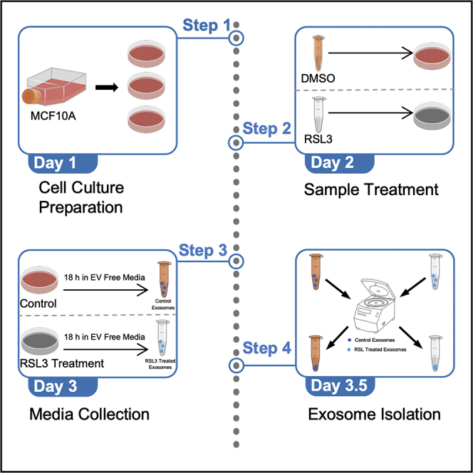
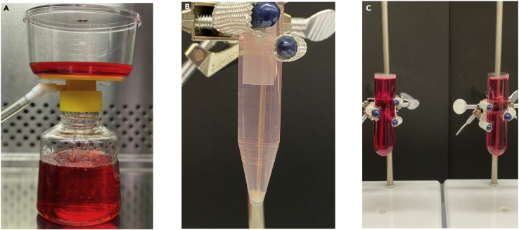
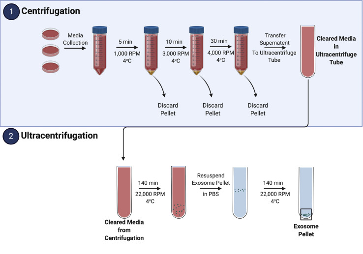
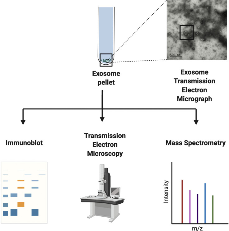
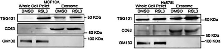
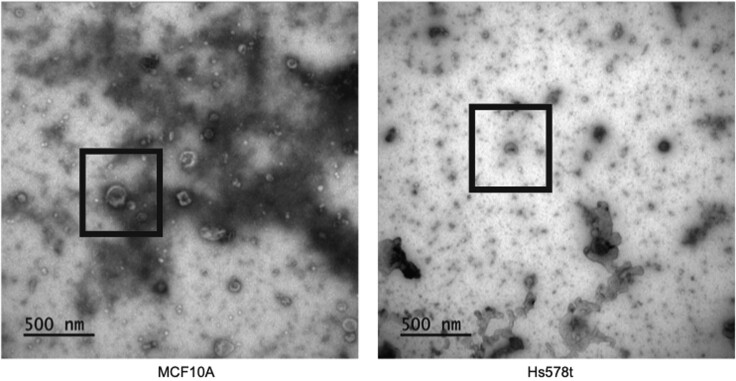
Extracellular vesicles (EVs) play key roles in transporting key molecular constituents as cargo for extracellular trafficking. While several approaches have been developed to extract EVs from mammalian cells, the specific method of EV isolation can have a profound effect on membrane integrity and yield. Here, we describe a step-by-step procedure to separate EVs from adherent epithelial cells using differential ultracentrifugation. Separated EVs can be further analyzed by immunoblotting, mass spectrometry, and transmission electron microscopy to derive EV yield and morphology. For complete details on the use and execution of this protocol, please refer to Brown et al. (2019).

Keywords: Cancer; Cell biology; Cell culture; Cell membrane.


Conflict of interest statement

The authors declare no competing interests.

Figures

None
Graphical abstract
Figure 1
Figure 1
Extracellular vesicle separation by ultracentrifugation step-by-step method overview (A) Media filter sterilization (0.22 μm filter) for subculturing cells. (B) MCF10A cell pellet after centrifugation at 3,000 rpm for 10 min at 4°C. (C) Two ultracentrifugation tubes balanced with media and using cold PBS as necessary. Ultracentrifuge tubes are filled to avoid collapse as a result of insufficient volume.
Figure 2
Figure 2
Illustrative extracellular vesicle isolation protocol overview After cell culture preparation and sample treatment, the media is collected and undergoes two phases for EV isolation and enrichment: (1) centrifugation and (2) ultracentrifugation. During (1), cells (alive and dead) and debris are removed. EVs are then separated and enriched in (2). Figure partially created using BioRender.
Figure 3
Figure 3
Methods of identification and validation of extracellular vesicles following isolation and enrichment via ultracentrifugation Following isolation and enrichment via ultracentrifugation, EV components can be further characterized using immunoblotting, TEM, and mass spectrometry. Figure partially created using BioRender and Brown et al., 2019.
Figure 4
Figure 4
Characterization of extracellular vesicle components by immunoblot Separated EVs from RSL3-treated MCF10A and Hs578t cells were immunoblotted for TSG101, GM130, and CD63. (Figure adapted from Brown et al., 2019).
Figure 5
Figure 5
TEM image of separated extracellular vesicles in MCF10A and Hs578t cells EVs separated from MCF10A and Hs578t cells were imaged by TEM. EVs are shown in black boxes. (Figure adapted from Brown et al., 2019).

References

    1. Bandu R., Oh J.W., Kim K.P. Mass spectrometry-based proteome profiling of extracellular vesicles and their roles in cancer biology. Exp. Mol. Med. 2019;51:1–10. - PMC - PubMed
    1. Brown C.W., Amante J.J., Chhoy P., Elaimy A.L., Liu H., Zhu L.J., Baer C.E., Dixon S.J., Mercurio A.M. Prominin2 drives ferroptosis resistance by stimulating iron export. Dev. Cell. 2019;51:575–586. - PMC - PubMed
    1. Cheng Y., Zeng Q., Han Q. Effect of pH, temperature and freezing-thawing on quantity changes and cellular uptake of exosomes. Protein Cell. 2019;10:295–299. - PMC - PubMed
    1. Coumans F.A.W., Brisson A.R., Buzas E.I., Dignat-George F., Drees E.E.E., El-Andaloussi S., Emanueli C., Gasecka A., Hendrix A., Hill A.F. Methodological guidelines to study extracellular vesicles. Circ. Res. 2017;120:1632–1648. - PubMed
    1. Cvjetkovic A., Lotvall J., Lasser C. The influence of rotor type and centrifugation time on the yield and purity of extracellular vesicles. J. Extracell. Vesicles. 2014;3 doi: 10.3402/jev.v3.23111. - DOI - PMC - PubMed

Publication types

MeSH terms

LinkOut - more resources