

Radiographic greater tuberosity spurs and narrow acromiohumeral intervals are associated with advanced retraction of the supraspinatus tendon in patients with symptomatic rotator cuff tears
- PMID: 33554169
- PMCID: PMC7846697
- DOI: 10.1016/j.jseint.2020.09.015
Radiographic greater tuberosity spurs and narrow acromiohumeral intervals are associated with advanced retraction of the supraspinatus tendon in patients with symptomatic rotator cuff tears
Abstract
Background: Degenerative signs on shoulder radiographs, including spur formation and narrow acromiohumeral intervals (AHIs), have been recognized as indicative of atrophic and fat-infiltrated rotator cuff muscles. Past studies have demonstrated that patients with poor quality muscles are prone to retraction of the supraspinatus tendon and failure to repair. However, the association between radiographic signs and tendon retraction has never been elucidated in previous literature. The present study aimed to investigate the association between the degenerative signs on shoulder radiographs and the severity of supraspinatus retraction.
Methods: Images of 67 individuals, who had undergone an arthroscopic rotator cuff repair, were retrospectively reviewed. The greater tuberosity (GT) morphology, subacromial spur, AHI, and acromial thickness were evaluated on the radiographs, whereas the retraction of the supraspinatus tendon was assessed via an MRI in accordance with the Patte classification. Simple regression analyses between the radiographic signs and Patte stages were performed, and factors reaching statistical significance were then included in the multiple ordinal logistic regression. Statistically significant predictors from the multiple regression analysis were constructed into combinations, for which the sensitivity and specificity were calculated.
Results: The GT morphology (P = .004), AHI (P = .083), subacromial spur (P = .008), and age (P = .004) were associated with supraspinatus retraction in the simple regression analyses. These four parameters were incorporated into the multiple ordinal logistic regression, where the GT spur (adjusted odds ratio 8.63, 95% confidence interval 2.16-34.53, P = .002) and AHI (AOR 0.79, 95% CI 0.63-0.98, P = .032) were demonstrated to be predictive of the Patte stage of supraspinatus retraction. The acromial spur implied a higher risk of severe retraction although this finding was not statistically significant (AOR 2.89, 95% CI 0.90-9.29, P = .075). The presence of concurrent GT spur and narrow AHI was highly specific (sensitivity 27.3% / specificity 91.1%) for advanced supraspinatus retraction.
Conclusion: The presence of a radiographic GT spur, narrow AHI, and subacromial spur indicated advanced retraction of the supraspinatus tendon. When patients with clinical suspicion of rotator cuff tear present with combinations of these radiographic signs, a prompt MRI examination and a referral to a shoulder specialist are recommended.
Keywords: Rotator cuff tear; greater tuberosity spur; radiography; tendon retraction.
© 2020 The Author(s).
Figures

 ); (C) Acromiohumeral interval (); (D) Normal GT; (E) Sclerotic GT (
 ); (F) Spurring GT (
 ). GT, greater tuberosity.
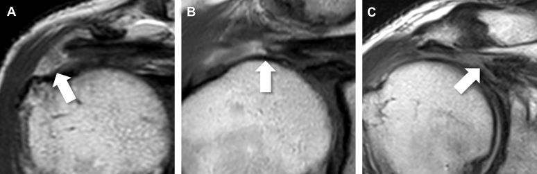
Similar articles
-
The radiographic morphology of the greater tuberosity is associated with muscle degeneration in patients with symptomatic rotator cuff tears.J Shoulder Elbow Surg. 2019 Oct;28(10):1964-1970. doi: 10.1016/j.jse.2019.03.010. Epub 2019 Jun 13. J Shoulder Elbow Surg. 2019. PMID: 31202626
-
Predictive Factors of Retear in Patients With Repaired Rotator Cuff Tear on Shoulder MRI.AJR Am J Roentgenol. 2018 Jan;210(1):134-141. doi: 10.2214/AJR.17.17915. Epub 2017 Oct 18. AJR Am J Roentgenol. 2018. PMID: 29045184
-
Relationship between the morphology of the greater tuberosity and radiological and clinical outcomes after arthroscopic rotator cuff repair.JSES Int. 2021 Jan 25;5(3):493-499. doi: 10.1016/j.jseint.2020.11.009. eCollection 2021 May. JSES Int. 2021. PMID: 34136860 Free PMC article.
-
A review of the role of magnetic resonance imaging in the evaluation of shoulder impingement syndrome and rotator cuff tendon tears.Ann Acad Med Singap. 1998 Mar;27(2):243-7. Ann Acad Med Singap. 1998. PMID: 9663318 Review.
-
US of the shoulder: rotator cuff and non-rotator cuff disorders.Radiographics. 2006 Jan-Feb;26(1):e23. doi: 10.1148/rg.e23. Radiographics. 2006. PMID: 16352733 Review.
Cited by
-
Transcending Patient Morphometry: Acromiohumeral Interval to Glenoid Ratio as a Universal Diagnostic Tool for Massive Rotator Cuff Tears.Clin Orthop Surg. 2024 Aug;16(4):578-585. doi: 10.4055/cios23381. Epub 2024 May 28. Clin Orthop Surg. 2024. PMID: 39092296 Free PMC article.
-
Superior humeral head osteophytes are associated with rotator cuff insufficiency in glenohumeral osteoarthritis: a retrospective analysis.Eur J Orthop Surg Traumatol. 2024 Feb;34(2):893-900. doi: 10.1007/s00590-023-03727-3. Epub 2023 Sep 28. Eur J Orthop Surg Traumatol. 2024. PMID: 37770594
-
Association Between Rotator Cuff Tears and Shoulder MRI Parameters: Importance of Arthroscopic Validation in Coronal Acromiohumeral Interval Measurement.Orthop J Sports Med. 2025 Jan 21;13(1):23259671241309695. doi: 10.1177/23259671241309695. eCollection 2025 Jan. Orthop J Sports Med. 2025. PMID: 39845420 Free PMC article.
References
-
- Bonsell S., Pearsall AWt, Heitman R.J., Helms C.A., Major N.M., Speer K.P. The relationship of age, gender, and degenerative changes observed on radiographs of the shoulder in asymptomatic individuals. J Bone Joint Surg Br. 2000;82:1135–1139. - PubMed
-
- Burns W.C., 2nd, Whipple T.L. Anatomic relationships in the shoulder impingement syndrome. Clin Orthop Relat Res. 1993;294:96–102. - PubMed
LinkOut - more resources
Full Text Sources
Other Literature Sources
Miscellaneous