. 2021 Jul;32(13-14):744-760.
doi: 10.1089/hum.2020.216. Epub 2021 May 5.

Characterization and Functional Analysis of CD44v6.CAR T Cells Endowed with a New Low-Affinity Nerve Growth Factor Receptor-Based Spacer

Affiliations

Characterization and Functional Analysis of CD44v6.CAR T Cells Endowed with a New Low-Affinity Nerve Growth Factor Receptor-Based Spacer

Anna Stornaiuolo et al. Hum Gene Ther. 2021 Jul.

Abstract

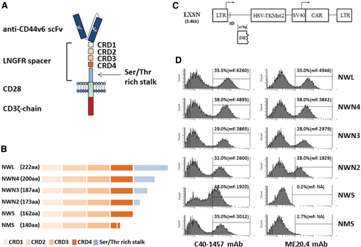
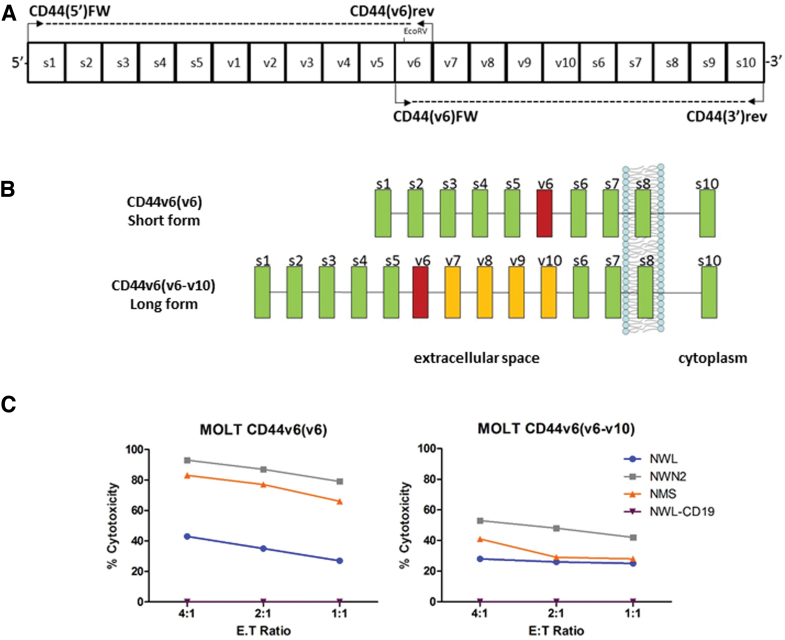
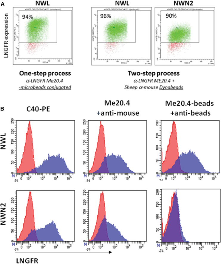
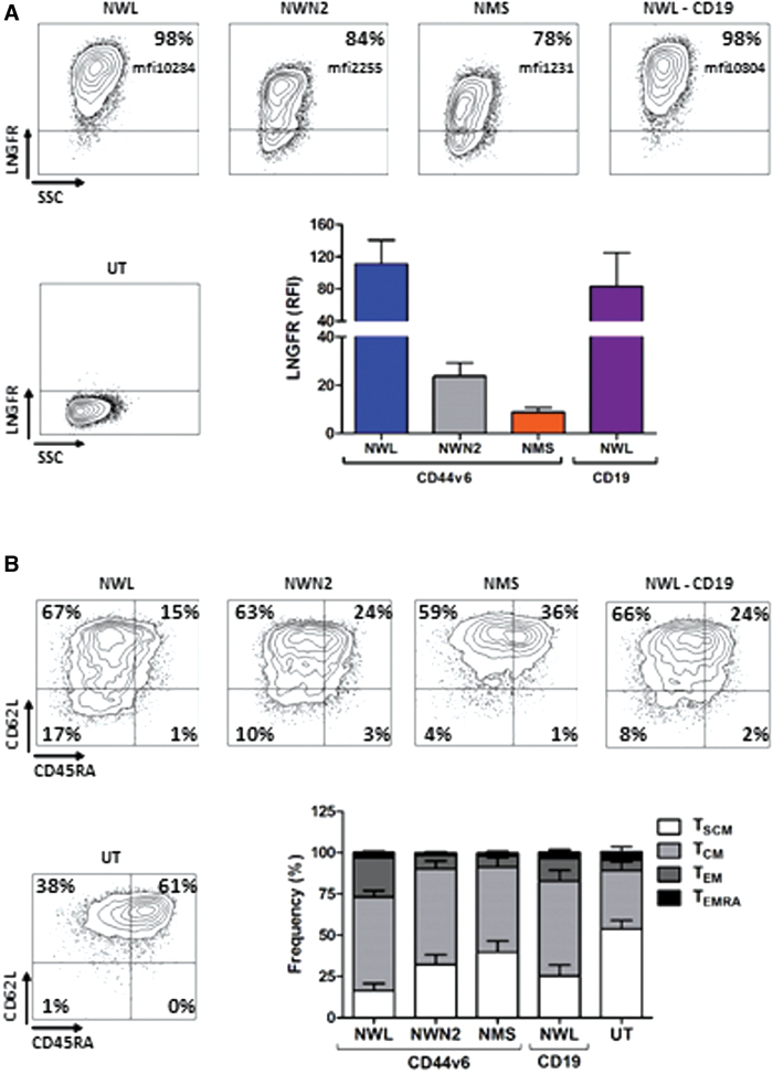
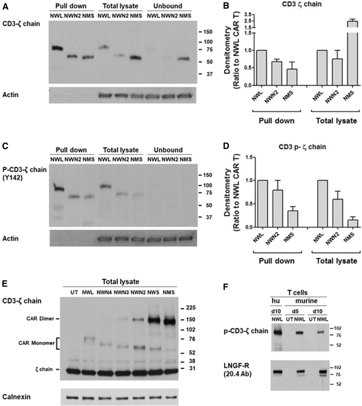
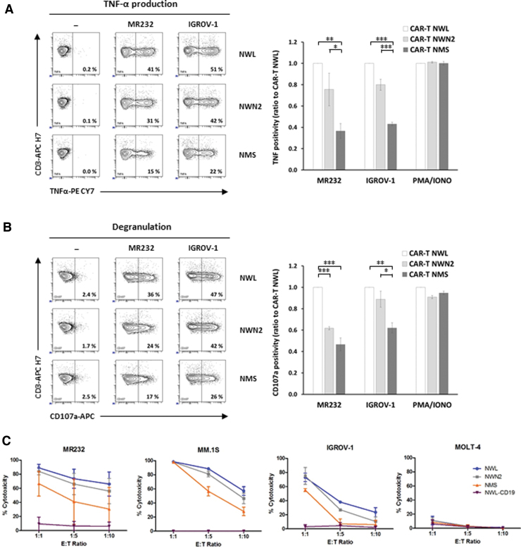
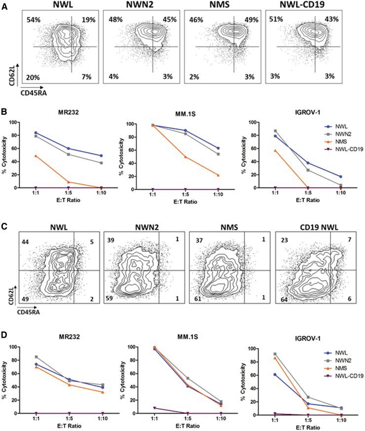
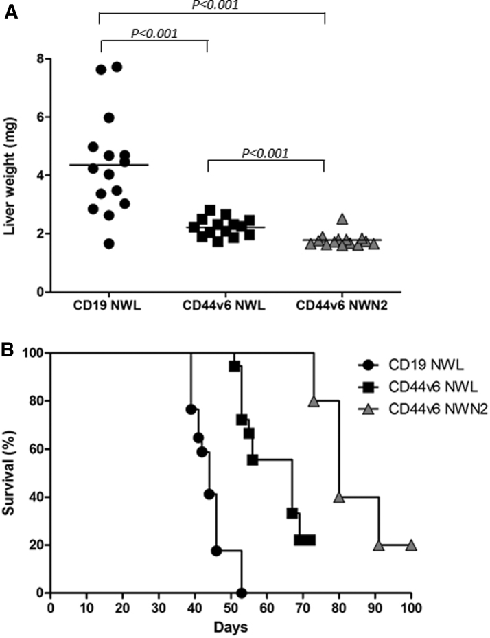
Effectiveness of adoptively transferred chimeric antigen receptor (CAR) T cells strongly depends on the quality of CAR-mediated interaction of the effector cells with the target antigen on tumor cells. A major role in this interaction is played by the affinity of the single-chain variable fragment (scFv) for the antigen, and by the CAR design. In particular, the spacer domain may impact on the CAR T cell function by affecting the length and flexibility of the resulting CAR. This study addresses the need to improve the manufacturing process and the antitumor activity of CD44v6-specific CAR T cells by defining the optimal structure of a spacer region derived from the extracellular domain of the human low-affinity nerve growth factor receptor (LNGFR). We tailored the LNGFR spacer to modulate CAR length to efficiently recognize distal or proximal epitopes and to allow selection of transduced CAR T cells by the use of clinical-grade validated manufacturing systems. The different LNGFR spacers investigated in this study are responsible for the generation of CAR T cells with a different memory phenotype, which is mainly related to the level of CAR expression and the extent of the associated tonic signaling. In particular, the CD44v6-NWN2.CAR T cells are enriched in central memory cells and show improved in vitro functions in terms of killing capability, and in vivo antitumor activity against hematological and solid tumors. Clinical Trial Registration numbers: clinicaltrial.gov NCT04097301; ClinicalTrials.gov, NCT00423124.

Keywords: CAR T; CD44v6; adoptive cell therapy; solid tumor; spacer.


Conflict of interest statement

C.S., C.A., and S.P. were MolMed employees. A.S., B.V., C.S., D.Z., Y.M.D.L.T., S.C., and C.T. are AGC Biologics SpA (formerly MolMed SpA) employees. AGC Biologics SpA (formerly MolMed SpA) is the applicant of patents on CAR molecules containing LNGFR-derived spacers including the CD44v6-targeted CARs studied in this work.

Figures

Figure 1.
Figure 1.
Features of the CAR constructs. (A) Schematic structure of the CD44v6-LNGFR CAR. The CAR contains a CD44v6 binding domain (anti-CD44v6 scFv), the LNGFR spacer (four TNFR-Cys domains [CRDs] and the serine/threonine-rich stalk), the transmembrane and costimulatory domain of CD28, and the intracellular domain of CD3ζ chain. (B) Schematic structure of the different LNGFR spacers. The LNGFR wild-type long spacer (NWL) contains the four CRD and the entire serine/threonine-rich stalk. The optimized LNGFR spacers N4 (NWN4), N3 (NWN3), and N2 (NWN2) contain the four CRDs and the first 38, 25, and 11 amino acids of the serine/threonine-rich stalk, respectively. The NWS spacer contains only the four CRDs, while the NMS spacer contains the first three CRDs and a deleted version of the CRD4. (C) Schematic representation of the retroviral vector construct LTK-SCD44v6-CAR derived from Moloney murine leukemia virus. The construct contains the transcriptional promoter 5′ viral long terminal repeat (5′LTR), the viral sequence including the packaging signal and gag gene (Ψ+ gag), the polynucleotide coding for the suicide gene HSV-TK Mut2, the transcriptional promoter SV40 (Simian virus 40), the CD44v6 CAR, and the 3′ viral long terminal repeat (3′LTR). (D) Characterization of the binding of the two anti LNGFR Abs, C40-1457 and ME20.4, to the CARs expressed by CEM A301 cells transduced with the different CAR constructs. Cells were analyzed at day 3 of culture. Percentage of transduction and MFI is shown in the inset. CAR, chimeric antigen receptor; LNGFR, low-affinity nerve growth factor receptor; MFI, median fluorescence intensity; scFv, single-chain variable fragment. Color images are available online.
Figure 7.
Figure 7.
Cloning of two CD44v6 isoforms. (A) Schematic representation of the coding region of human CD44 gene (s1-s10: standard exon; v1-v10: variant exon) and localization of the primers used to amplify the CD44 transcripts. (B) Schematic representation of CD44 and CD44v6 cloned isoforms: CD44v6(v6) from activated T lymphocytes, CD44v6(v6-v10) from THP-1. Box color code: green indicates standard exons; yellow indicates variable exons; magenta indicates variable exon 6. (C) Cytotoxic activity of the differentiated CAR T cells was assessed by coculture with MOLT-CD44v6(v6) or MOLT-CD44v6(v6-v10), at the indicated E:T ratio. Results obtained from a representative experiment out of two from independent donors are shown (mean ± SEM). Color images are available online.
Figure 2.
Figure 2.
Immune-selection of CAR T cells. (A) CAR T cells endowed with NWL and NWN2 spacers were immune-selected by the use of either a single-step protocol with the mAb ME20.4 coupled to magnetic MicroBeads (CliniMACS CD271-LS MicroBeads; Miltenyi), or by a two-step method using mAb ME20.4 followed by selection with beads conjugated to anti-mouse antibodies (Dynabeads; Dynal). Both immunoselection processes allowed the isolation of highly enriched (>90%) populations of CAR T cells endowed with NWL spacer, whereas CD44v6-NWN2.CAR T were purified only with the two-step method. (B) Pure populations of CAR T cells expressing the NWL or the NWN2 CAR constructs were stained with (1) the anti-LNGFR mAbs C40-PE (left panels); (2) the ME20.4 mAb followed by an anti-mouse-PE (middle panels); and (3) the ME20.4 mAb coupled to MicroBeads followed by an anti-MicroBead-PE (right panels). NWL expressing CAR T cells are recognized by all the combinations tested (upper panels). Concerning the NWN2 construct (lower panels), ME20.4 mAb is able to bind the transduced cells and can be in turn recognized by an anti-mouse mAb, thus justifying the successful selection obtained with the two-step procedure. On the contrary, when directly coupled to the MicroBeads, the ME20.4 mAb is no more able to bind the NWN2.CAR. mAb, monoclonal antibody. Color images are available online.
Figure 3.
Figure 3.
Characterization of CD44v6.CAR T cells used for functional analysis. (A) CAR expression in transduced T lymphocytes detected with the anti-LNGFR mAb C40-1457. Plots of a representative experiment are shown on the left. On the right, means with SDs of the results obtained with eight independent donors are shown. Paired t-test on LNGFR expression was statistically significant for CD44v6-NWN2 (p ≤ 0.05) and CD44v6-NMS (p ≤ 0.05) compared with CD44v6-NWL cells. (B) T cell memory phenotype was defined based on CD45RA and CD62L expression at day 10 after transduction. Plots of a representative donor are shown on the left. On the right, means with SDs of the results obtained with eight independent donors are shown. Paired t-test on TSCM was statistically significant for untransduced (UT; p ≤ 0.0001), CD44v6-NWN2 (p ≤ 0.001), CD44v6-NMS (p ≤ 0.001), and CD19-NWL (p ≤ 0.01) cells, compared with CD44v6-NWL cells. TSCM (CD45RA+/CD62L+ memory stem T cells), TCM (CD45RA/CD62L+ central memory T cells), TEM (CD45RA/CD62L effector memory T cells), TEMRA (CD45+/CD62L effector memory RA T cells). Color images are available online.
Figure 4.
Figure 4.
Biochemical analysis of the CD44v6.CAR expression and phosphorylation. (A) Samples of cell surface protein (pull-down), total lysates, or unbound fractions, from the different CD44v6.CAR T, were immunoblotted with the CD3-ζ chain Ab, or actin as loading control. A representative experiment out of two is shown. (B) Densitometric analysis of the bands shown in A from two experiments performed with different donors was performed and results were expressed as ratio to the CD44v6-NWL.CAR T (mean ± SE). (C) Samples of cell surface protein (pull-down), total lysates, or unbound fractions, from the different CD44v6.CAR T, were immunoblotted with the anti-phospho-CD3-ζ chain (Tyr142) Ab, or actin as loading control. A representative experiment out of three is shown. (D) Densitometric analysis of the bands shown in C from two experiments performed with different donors was performed and results were expressed as ratio to the CD44v6-NWL.CAR T (mean ± SE). (E) Total lysates from CD44v6.CAR T cells were analyzed by immunoblotting with the anti CD3-ζ chain Ab in nonreducing conditions, and normalization was performed on the same filter by calnexin blot. A representative experiment out of three is shown. (F) Total lysates from murine (at day 10 or 15 of culture) or human (hu; day 10) CD44v6-NWL.CAR T cells were analyzed by immunoblotting with the anti-phospho-CD3-ζ chain (Tyr142) Ab. Normalization was obtained by LNGFR immunoblotting. A representative experiment out of two is shown.
Figure 5.
Figure 5.
Functional analysis of CD44v6 CAR T cells in vitro. (A, B) Activation of the three CD44v6.CAR T cells (NWL, NWN2, and NMS) was analyzed by coculture with MR232, IGROV-1 (E:T ratio 1:1), or after PMA/ionomycin stimulation (5 h of incubation). Percentage of TNF-α-positive (A) or CD107a-positive (B) CAR T cells was analyzed by FACS analysis (representative plots on the left). The results obtained from three independent donors are shown (mean ± SEM) in the graphs, expressed as relative ratio to CD44v6-NWL.CAR T positivity. *p < 0.05, **p < 0.01, ***p < 0.001 (Bonferroni's multiple comparison test). (C) Cytotoxic activity of the three CD44v6.CAR T cells (NWL, NWN2, and NMS) and the CD19-NWL.CAR T cells was analyzed by coculture with the indicated target cells, expressing luciferase, at different ratios. After 24 h, the bioluminescence emitted by the live target cells was detected. The results obtained from two independent donors are shown (mean ± SEM). PMA, phorbol myristate acetate. Color images are available online.
Figure 6.
Figure 6.
Differentiation status influences the cytotoxic activity of CD44v6.CAR T cells. CD44v6.CAR T cells (NWL, NWN2, and NMS) and CD19-NWL.CAR T cells were kept early differentiated (A) or in vitro differentiated (C) with IL2 (100 U/mL) and OKT-3 (30 ng/mL), and their memory phenotype was defined by CD45RA and CD62L expression. TSCM (CD45RA+/CD62L+ memory stem T cells), TCM (CD45RA/CD62L+ central memory T cells), TEM (CD45RA/CD62L effector memory T cells), TEMRA (CD45+/CD62L terminally differentiated T cells). Results obtained from a representative donor are shown. Cytotoxic activity of the early differentiated (B) or differentiated (D) CAR T cells was analyzed after 24 h of coculture with the MR232, MM.1S, and IGROV-1 target cells at the indicated E:T ratio. Results obtained from a representative experiment out of two from independent donors are shown. Color images are available online.
Figure 8.
Figure 8.
In vivo antitumor activity. (A) CD44v6 CAR T cell-mediated antileukemic effect was evaluated in the well-established THP-1 high-burden disease model. Liver weight (mg) in the different treatment groups at sacrifice (6 weeks) is shown (n = 3 independent experiments with different T cell donors). Results from paired T test are shown. (B) NSG mice were subcutaneously injected with 3 × 105 CD44v6+ IGROV-1 tumor cells. Seven days later, mice were infused via tail vein injection with 4.5 × 106 CD19 or CD44v6.CAR T cells and tumor growth quantified by measuring tumor size. Mice treated with CD44v6-NWN2.CAR T cells showed an enhancement of survival several weeks after treatment compared with CD19-NWL.CAR T mice (n = 17; p ≤ 0.0002), as well as with CD44v6-NWL.CAR T (n = 18; p ≤ 0.0075). Results from log-rank test are reported.

References

    1. Sadelain M, Riviere I, Brentjens R. Targeting tumours with genetically enhanced T lymphocytes. Nat Rev Cancer 2003;3:35–45 - PubMed
    1. Ho WY, Blattman JN, Dossett ML, et al. . Adoptive immunotherapy: engineering T cell responses as biologic weapons for tumor mass destruction. Cancer Cell 2003;3:431–437 - PubMed
    1. Jafarzadeh L, Masoumi E, Fallah-Mehrjardi K, et al. . Prolonged persistence of chimeric antigen receptor (CAR) T cell in adoptive cancer immunotherapy: challenges and ways forward. Front Immunol 2020;11:702. - PMC - PubMed
    1. Sabatino M, Hu J, Sommariva M, et al. . Generation of clinical-grade CD19-specific CAR-modified CD8+ memory stem cells for the treatment of human B-cell malignancies. Blood 2016;128:519–528 - PMC - PubMed
    1. van der Stegen SJ, Hamieh M, Sadelain M. The pharmacology of second-generation chimeric antigen receptors. Nat Rev Drug Discov 2015;14:499–509 - PMC - PubMed

Publication types

Substances

Associated data