. 2021 Mar 1;320(3):F454-F463.
doi: 10.1152/ajprenal.00426.2020. Epub 2021 Feb 8.

Mesenchymal stem cells protect renal tubular cells via TSG-6 regulating macrophage function and phenotype switching

Affiliations

Mesenchymal stem cells protect renal tubular cells via TSG-6 regulating macrophage function and phenotype switching

Yu Zhao et al. Am J Physiol Renal Physiol. .

Abstract

Tumor necrosis factor (TNF)-α-induced gene/protein (TSG)-6 regulates the immunomodulatory properties of mesenchymal stem cells (MSCs), but its ability to protect the ischemic kidney is unknown. In a swine model of renal artery stenosis (RAS) and metabolic syndrome (MetS), we assessed the contribution of TSG-6 produced by MSCs to their immunomodulatory properties. Pigs were studied after 16 wk of diet-induced MetS and unilateral RAS and were either untreated or treated 4 wk earlier with intrarenal autologous adipose tissue-derived MSCs (n = 6 each). Lean, MetS, and RAS sham animals served as controls. We studied renal function in vivo (using computed tomography) and kidney histopathology and macrophage phenotype ex vivo. In vitro, TSG-6 levels were also measured in conditioned media of human MSCs incubated with TNF-α and levels of the tubular injury marker lactate dehydrogenase in conditioned media after coculturing macrophages with injured human kidney 2 (HK-2) cells with or without TSG-6. The effects of TSG-6 on macrophage phenotype (M1/M2), adhesion, and migration were also determined. MetS + RAS showed increased M1 macrophages and renal vein TNF-α levels. After MSC delivery, renal vein TSG-6 increased and TNF-α decreased, the M1-to-M2 ratio decreased, renal function improved, and fibrosis was alleviated. In vitro, TNF-α increased TSG-6 secretion by human MSCs. TSG-6 decreased lactate dehydrogenase release from injured HK-2 cells, increased expression of macrophage M2 markers, and reduced M1 macrophage adhesion and migration. Therefore, TSG-6 released from MSCs may decrease renal tubular cell injury, which is associated with regulating macrophage function and phenotype. These observations suggest that TSG-6 is endowed with renoprotective properties.NEW & NOTEWORTHY Tumor necrosis factor-α-induced gene/protein (TSG)-6 regulates the immunomodulatory properties of MSCs, but its ability to protect the ischemic kidney is unknown. In pigs with renal artery stenosis, we show that MSC delivery increased renal vein TSG-6, decreased kidney inflammatory macrophages, and improved renal function. In vitro, TSG-6 decreased inflammatory macrophages and tubular cell injury. Therefore, TSG-6 released from MSCs may decrease renal tubular cell injury, which is associated with regulating macrophage function and phenotype.

Keywords: macrophage; mesenchymal stem cells; metabolic syndrome; renal artery stenosis; tumor necrosis factor-α-induced gene/protein-6.


Figures

None
Graphical abstract
Figure 1.
Figure 1.
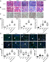
Pig mesenchymal stem cell (p-MSC) characterization and tracking. A: schematic of the experimental protocol. B: images of CM-DiI-labeled (red) p-MSCs in frozen sections of the poststenotic kidney of pigs from the metabolic syndrome (MetS) + renal artery stenosis (RAS) group 4 wk after cell delivery. C: in frozen sections of MetS + RAS kidneys that received MSCs, immunofluorescent staining with cytokeratin staining identified MSCs within tubular cells. D: lactate dehydrogenase (LDH) levels in urine. Results are means ± SD (t test). *P < 0.05 vs. the lean group (n =5 or 6 pigs/group).
Figure 2.
Figure 2.
Pig mesenchymal stem cells (p-MSCs) induce a shift in renal pathology and macrophage phenotype. At the end of the study, kidneys were harvested, paraffin sections were used for Masson’s trichrome staining (A and B), and semiquantitative analysis of the renal cortex (D) and medulla (E) demonstrated renal fibrosis in the metabolic syndrome (MetS) + renal artery stenosis (RAS) group that decreased after p-MSC treatment. Periodic acid-Schiff (PAS) stains showed a similar trend for renal tubular injury (C and F). G–J: representative merged images (×40) of immunofluorescence staining for stenotic kidney M1 macrophages [CD68 (red)/inducible nitric oxide synthase (green)] and M2 macrophages [CD68/arginase-1 (green)] (double staining yellow). Quantification of stenotic kidney M1 and M2 macrophages and the M1-to-M2 ratio is shown. Results are means ± SD (t test). *P < 0.05 vs. the lean group; #P < 0.05 vs. the MetS + RAS group; †P < 0.05 vs. the MetS group; &P < 0.05 vs. the RAS group (n =5 or 6 pigs/group).
Figure 3.
Figure 3.
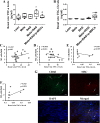
Link between tumor necrosis factor (TNF)-α and the macrophage M1/M2 phenotype. Renal vein plasma samples were collected using a catheter at the end of the study, and levels of TNF-α (A) and TNF-α-induced gene/protein (TSG-6; B) were determined. Renal vein TNF-α correlated directly with M1 (C) and inversely with M2 (D) macrophage numbers. Renal vein TSG-6 levels showed no correlation with M1 macrophages (E) but correlated directly with M2 macrophages (F). G: in frozen sections of metabolic syndrome (MetS) + renal artery stenosis (RAS) kidneys treated with pig mesenchymal stem cells (p-MSCs), p-MSCs (red) incorporated within kidney macrophages (green). Results are means ± SD (t test). *P < 0.05 vs. the lean group; #P < 0.05 vs. the MetS + RAS group; †P < 0.05 vs. the MetS group; &P < 0.05 vs. the RAS group (n =5 or 6 pigs/group, except for E and F, where n =6 pigs each in the MetS + RAS and MetS + RAS + p-MSC groups).
Figure 4.
Figure 4.
Tumor necrosis factor (TNF)-α increased TNF-α-induced gene/protein (TSG-6) expression, which reduced renal tubular cell injury. A: representative Western blot of TSG-6 protein expression in TNF-α-stimulated human mesenchymal stem cells (h-MSCs) harvested from healthy human subjects. GAPDH was used as an internal control. B: supernatant TSG-6 levels in TNF-α-induced h-MSCs. Human tubular cell line HK-2 cell vitality fell time dependently by TNF-α and antimycin-A (AMA) treatment (C), whereas lactate dehydrogenase (LDH) levels rose (D). E and F: supernatant TNF-α (E) and LDH (F) levels after TSG-6 stimulation of M1 macrophages. Results are means ± SE for experiments performed in triplicate (t test). *P < 0.05 vs. the normal control (NC); #P < 0.05 vs. TNF-α + AMA.
Figure 5.
Figure 5.
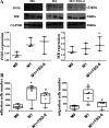
Tumor necrosis factor-α-induced gene/protein (TSG-6) attenuates M1/M2 macrophage phenotype, adhesion, and migration. Human monocytes (U937 cells) were stimulated and polarized toward M1 macrophages and then treated with TSG-6 in vitro. A: TSG-6 increased M2 marker mannose receptor (MR) expression in macrophages (M0) (GAPDH loading control). B: TSG-6 decreased M1 macrophage adhesion and migration. iNOS, inducible nitric oxide synthase. Results are means ± SE for experiments performed in triplicate (t test). *P < 0.05 vs M0; #P < 0.05 vs M1.
Figure 6.
Figure 6.
Illustration of the renoprotective properties of mesenchymal stem cells (MSCs) potentially mediated through tumor necrosis factor-α-induced gene/protein (TSG-6) regulation of macrophages. TNF-α-induced TSG-6 release from MSCs may decrease renal tubular cells injury, which is associated with, and may be partly mediated by, inducing a macrophage phenotypic switch from M1 to M2 macrophages and reducing M1 macrophage adhesion and migration.

References

    1. Eirin A, Zhu XY, Jonnada S, Lerman A, van Wijnen AJ, Lerman LO. Mesenchymal stem cell-derived extracellular vesicles improve the renal microvasculature in metabolic renovascular disease in swine. Cell Transplant 27: 1080–1095, 2018. doi:10.1177/0963689718780942. - DOI - PMC - PubMed
    1. Zhu XY, Urbieta-Caceres V, Krier JD, Textor SC, Lerman A, Lerman LO. Mesenchymal stem cells and endothelial progenitor cells decrease renal injury in experimental swine renal artery stenosis through different mechanisms. Stem Cells 31: 117–125, 2013. doi:10.1002/stem.1263. - DOI - PMC - PubMed
    1. Zhuang Q, Ma R, Yin Y, Lan T, Yu M, Ming Y. Mesenchymal stem cells in renal fibrosis: the flame of cytotherapy. Stem Cells Int 2019: 8387350, 2019. doi:10.1155/2019/8387350. - DOI - PMC - PubMed
    1. Ebrahimi B, Eirin A, Li Z, Zhu XY, Zhang X, Lerman A, Textor SC, Lerman LO. Mesenchymal stem cells improve medullary inflammation and fibrosis after revascularization of swine atherosclerotic renal artery stenosis. PLoS One 8: e67474, 2013. doi:10.1371/journal.pone.0067474. - DOI - PMC - PubMed
    1. Choi H, Lee RH, Bazhanov N, Oh JY, Prockop DJ. Anti-inflammatory protein TSG-6 secreted by activated MSCs attenuates zymosan-induced mouse peritonitis by decreasing TLR2/NF-kappaB signaling in resident macrophages. Blood 118: 330–338, 2011. doi:10.1182/blood-2010-12-327353. - DOI - PMC - PubMed

Publication types

LinkOut - more resources